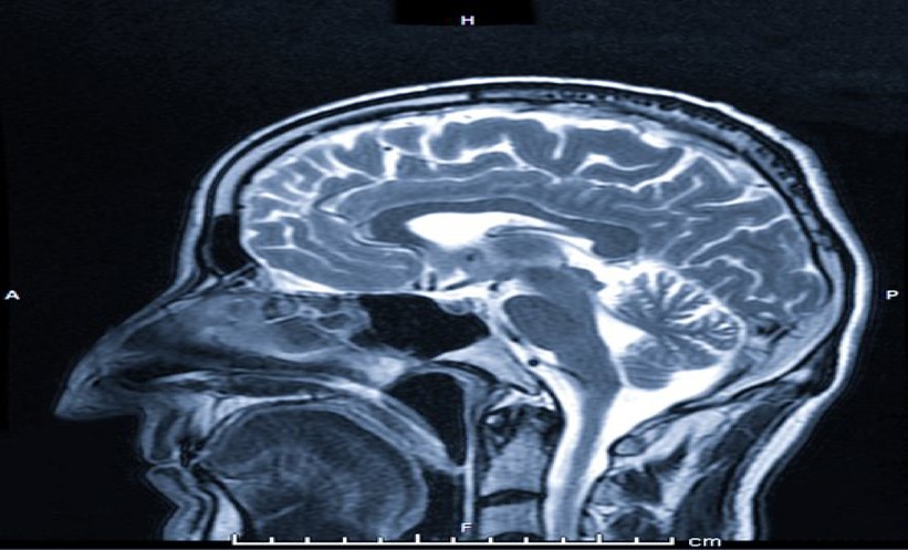
In the high-stakes environment of emergency neurology, clinicians often have mere minutes to predict the trajectory of a patient suffering from a primary brainstem haemorrhage.
The research, which examined 372 patients, highlights a shift toward using objective, computer-generated data to supplement the subjective interpretation of emergency scans. By utilizing a three-dimensional U-Net for haematoma segmentation, researchers developed a framework that provides a more granular look at the damage caused by these severe strokes. The findings suggest that this technology could serve as a vital tool for early risk stratification.
Predicting whether a patient will face poor functional outcomes at discharge is notoriously difficult. The study, authored by Y. Li and colleagues, found that their combined model—which integrates automated radiomics features with traditional radiological predictors—achieved an area under the curve (AUC) of 0.926 in the training dataset and 0.858 in the temporal internal validation dataset. These figures represent a notable improvement over standard assessment methods.
The researchers said that the automated segmentation performed comparably to the manual segmentation. By removing the need for time-consuming manual intervention, the system could theoretically be integrated into existing emergency workflows, providing physicians with rapid, data-driven insights. Using SHapley Additive exPlanations (SHAP), the team identified that the classification of the haematoma itself was the most influential predictor in the prognostic model.
While the results for functional outcomes were promising, the model’s ability to predict in-hospital post-stroke pneumonia (PSP) remained inconsistent. The combined PSP model saw AUCs drop to 0.681 in the validation dataset. Further refinement of the algorithm is clearly necessary before it can be used to reliably flag infection risks.
For those working in acute stroke care, the gap between a promising research model and a reliable clinical tool is vast. The study emphasizes that while the automated system is feasible, it is meant to act as a support mechanism. The primary challenge remains the validation of these models.
The stakes for the medical community are high. Brainstem haemorrhages are among the most devastating forms of stroke, frequently leading to profound disability or death. Current prognostic tools often rely on static clinical scores that may not capture the nuances of structural brain changes, leaving families and clinicians in a state of uncertainty regarding long-term recovery.
The use of SHAP analysis in this study is a step toward transparency, allowing clinicians to see exactly which features of the brain scan are driving the model’s predictions. This transparency is essential for building trust in diagnostic tools that will eventually inform life-altering decisions.
As the medical field continues to move toward more precise, data-heavy interventions, the integration of radiomics into standard stroke protocols remains a work in progress. Researchers continue to refine the tools required to manage one of the most complex medical emergencies in modern practice, but it is unclear if these models can be scaled to the bedside.
Worth a look

Sunflower Children’s Hospice Construction to Begin in Castlebar
Construction work on the first children’s hospice outside Dublin is expected to begin in January 2027, changing how...

Samsung Galaxy A18 5G Surfaces in Bluetooth Database
Samsung is preparing to expand its device lineup with the upcoming Galaxy A18 5G, which has surfaced on...
Broad Genomic Sequencing Extends Advanced Lung Cancer Survival
Broad genomic sequencing extended overall survival in advanced non-small cell lung cancer patients by an average of two...

Tulfo Files Resolution Seeking Senate Probe Into Spaghetti Wires
Senator Raffy Tulfo filed a resolution seeking a Senate investigation into hazardous overhead utility wires after a series...