
Researchers analyzing pediatric infective endocarditis find that prosthetic valve infections occur at an older age and show lower echocardiography sensitivity than native valve cases, requiring broader multimodality imaging and frequent surgical intervention, according to a study published on September 2, 2026.
Clinical Differences Between Native and Prosthetic Valve Infections in Children
Infective endocarditis in pediatric patients presents distinct clinical courses depending on whether the infection involves native heart tissue or artificial material. A retrospective review of clinical records from a specialised paediatric cardiac centre in Italy examined outcomes across 95 consecutive patients younger than 18 years (median age at diagnosis, 14 years; 62.2% boys) who met the modified Duke criteria for definite or possible infective endocarditis between 2013 and 2024.
The study divided the cohort into two groups: patients with native valve endocarditis (NVE) numbering 30, and those with prosthetic valve endocarditis (PVE) accounting for 65 participants. Researchers found that patients dealing with artificial valve infections were significantly older at diagnosis than those with native valve infections, with a median age of 15.7 years compared to 7.8 years (P = .00002).
Congenital heart disease appeared in 88.4% of the overall patient group. When looking at specific heart structures, pulmonary valve involvement proved significantly more common in prosthetic infections than in native ones at 64.4% versus 23.3% (P = .0002). Conversely, leaflet perforation appeared much more frequently in native valve endocarditis at 30.0% compared to just 1.5% in prosthetic cases (P = .00002).
Pathogen Profiles and Diagnostic Challenges on Echocardiography
Bacterial causes varied across the two clinical groups. Staphylococcus aureus served as the most frequent pathogen in native valve infections, accounting for 30.4% of positive cultures. In contrast, viridans streptococci predominated in prosthetic valve cases at 31.1% of positive cultures.
Coagulase-negative staphylococci appeared exclusively in prosthetic valve infections at 23.4% compared to zero percent in native cases (P = .01), although overall pathogen distributions did not show a statistically significant difference between the two categories. Blood cultures returned negative results in 28.4% of patients, a factor researchers attributed to potential referral after antimicrobial therapy had already started.
Detecting vegetations and lesions posed a distinct hurdle on standard ultrasound equipment. Echocardiography identified lesions suspicious for endocarditis less frequently in prosthetic cases at 67.7% compared to 93.3% in native valve infections (P = .007). This limitation drove a heavier reliance on advanced imaging techniques.
Clinicians turned to cardiac computed tomography angiography (CTA) for 55.4% of prosthetic valve patients versus 26.7% of native valve patients (P = .01), while metabolic imaging (PET/SPECT) was utilized in 35.4% of prosthetic cases compared to 13.3% of native cases (P = .03).
“The presence of IE [infective endocarditis] involving prosthetic material dictates a specialized diagnostic approach utilizing multimodality imaging, including CTA [CT angiography] and metabolic studies (PET/SPECT [single-photon emission CT]), to reliably confirm infection in cases where echocardiography sensitivity is compromised, or when additional information is required, as well as a multidisciplinary team management mandated by modern ESC [European Society of Cardiology] guidelines,”
the authors of the study wrote.
Complications, Surgical Intervention, and Follow-Up Outcomes
Complicated infective endocarditis struck 73.7% of the total cohort, defined by the presence of heart failure, systemic or pulmonary thromboembolic events, organ injury such as kidney damage or neurologic deficit, or local valvular, perivalvular, or periprosthetic complications.
More than half of the patients, specifically 54.7%, required cardiac surgery. Procedures were performed primarily to address heart failure in 48% of those surgical cases, uncontrolled infection in 27%, or thromboembolism in 25%. In-hospital mortality for the study population stood at 5.3%.
During subsequent follow-up periods, six patients experienced a recurrence of infective endocarditis, though only one patient suffered a recurrence involving the same pathogen within a 12-month window. The study was led by R. Iacobelli from Pediatric Cardiology, Clinical Area of Fetal and Cardiovascular Sciences, Bambino Gesù Children’s Hospital, IRCCS, Rome, Italy. It was supported by the Italian Ministry of Health through Current Research Funds
and published online on September 02, 2026, in the European Journal of Pediatrics.
Worth a look

Troponin Elevation in 20.4% of NSTE-ACS Patients
Out of 481 patients with non–ST-segment elevation acute coronary syndrome, 98 individuals—representing 20.4% of the cohort—met angiographic criteria...

Nitrous Oxide Abuse Linked to Rhabdomyolysis and AKI
A 22-year-old man developed severe rhabdomyolysis.
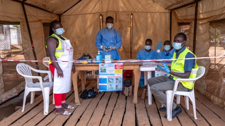
Ebola Outbreaks Linked to Distant Macroclimatic Shifts by Study
Distant macroclimatic oscillations modify continental rainfall and alter ecological conditions to create seasonal windows that trigger Ebola virus...

High-Nicotine Vapes Linked to Greater Nicotine Dependence in Young Adults
Between 2017 and 2022, high-nicotine e-cigarette use surged nearly 80 percent among young adults aged 18 to 24...