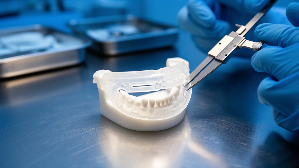
Children with severe temporomandibular joint ankylosis achieved immediate jaw openings exceeding 30 millimeters after surgery guided by patient-specific, 3D-printed arthroplasty guides, a prospective study at Alexandria University Teaching Hospital found.
The pilot investigation, published in Maxillofacial Plastic and Reconstructive Surgery, enrolled seven patients under age 15 between October 2024 and October 2025, according to Bioengineer. All presented with advanced intra-articular bony ankylosis classified as type III-A3 in five joints and type IV-A4 in two joints, and a mean preoperative maximal interincisal opening of just 9.41 mm.
Virtual surgical planning used high-resolution CT scans processed through Mimics segmentation software to construct 3D craniofacial models. Custom guides were CAD/CAM-fabricated with 5-mm-wide osteotomy slits designed to dictate cut direction and paired with ultrasonic piezoelectric bone cutting, the journal article reports.
Quantitative comparison of preoperative plans with postoperative CT scans showed a surgical angular deviation of 2.36 ± 0.18° on the right side and 1.90 ± 0.46° on the left. Linear vertical ramus height deviation measured 1.57 ± 0.33 mm on the right and 0.95 ± 0.46 mm on the left, demonstrating “good agreement between planned and postoperative measurements for both parameters,” the study authors wrote.
No intraoperative injury to the skull base or neurovascular structures occurred. One patient experienced temporary facial nerve weakness that resolved within eight weeks, according to the Europe PMC version of the article. The trial was retrospectively registered at ClinicalTrials.gov (NCT07320911).
Researchers concluded that the direction-dictating guides enable accurate and safe gap arthroplasty in pediatric patients, potentially reducing surgical variability. The study was conducted under PROCESS guidelines with institutional ethics approval and guardian consent.

Metropolitan Community College Sets $72 Flat Tuition for All Students Dec. 7
Metropolitan Community College in Omaha will charge the same $72 per credit hour to every student starting Dec....

Parents Coalition, Two Lawmakers Draft Bill for 45-Minute Recess
Elementary school students in Nevada could soon be guaranteed at least 45 minutes of daily recess under a...
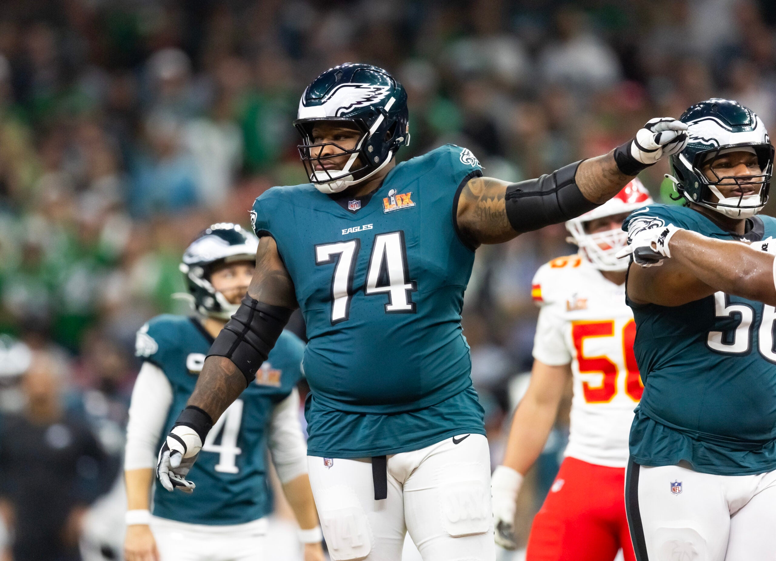
Chiefs Sign Former Eagles Lineman Fred Johnson to Practice Squad Amid Injuries
The Chiefs lost Super Bowl LIX to Philadelphia, then watched the Eagles cut the veteran lineman who helped...
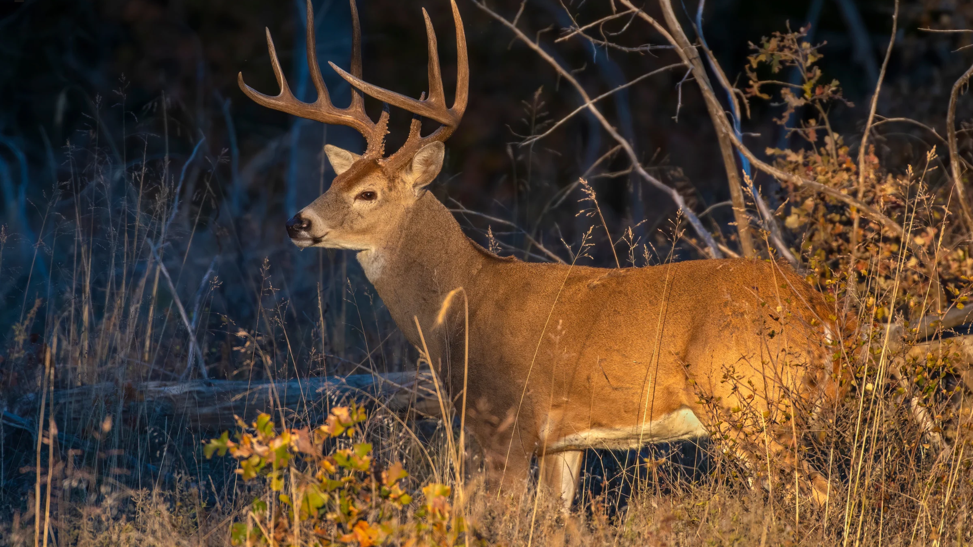
Mississippi Inspection Finds Violations at 10 Deer Breeding Pens; Duff, Deviney Facilities Cited
Mississippi wildlife inspectors found widespread record-keeping and sampling failures at 10 of 12 high-fence deer breeding pens this...