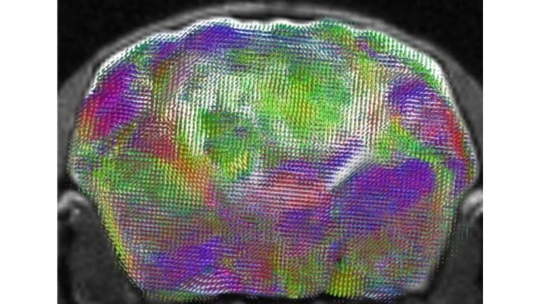
Stanford Medicine researchers have successfully transplanted lab-grown human cortical organoids into bioengineered mice missing most of their cerebral cortex, creating a living model where human nerve cells expanded to fill over 90 percent of the region and formed working neural connections, as detailed in research published September 16 in Nature.
Scientists have long chased a reliable way to study the human brain outside the human body. Because living human tissue is almost always inaccessible, researchers have long relied on cell cultures in laboratory glassware. While those miniature brain structures, known as organoids, have taught researchers a great deal since they first emerged in the early 2010s, they lacked the sensory inputs and complex connections of a living nervous system. Now, a team at Stanford Medicine has bridged that gap by placing lab-grown human brain tissue directly into the brains of living rodents.
Researchers transplanted self-organizing clusters of human cortical tissue into newborn mice that had been genetically engineered to develop without most of their cerebral cortex. The resulting cavity provided a hospitable environment where the human tissue could survive, thrive, and wire itself into the host animal.
Overcoming the Speed Barrier of Human Brain Development
Earlier attempts to install human brain cells into rodents repeatedly ran into an immovable biological obstacle. Human brain tissue develops at least 20 times slower than mouse brain tissue. In standard animal models, native mouse neurons formed rapid networks that quickly outcompeted the slower-developing human cells, closing the window of brain development before the human tissue could establish meaningful connections.
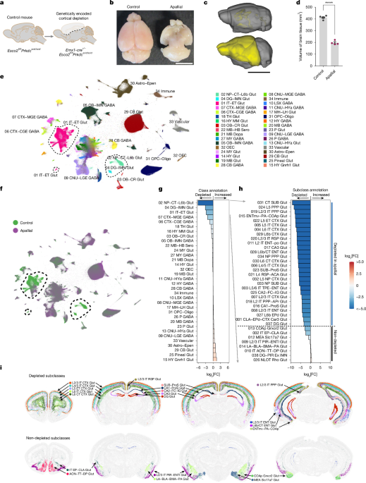
To solve this, the research team bred genetically engineered mice that were missing specific parts of their brains. By cleanly removing those structures ahead of time, the scientists left a vacant space that gave the incoming human cells ample room to grow without being immediately overwhelmed by native circuitry.

Surprisingly, the depleted mice remained functional. Once the pea-sized clumps of human neurons were placed into that vacant space, they quickly took root.
“Within a few days, the cell starts to divide and expand. Within a few weeks to months, they will essentially grow, become vascularized, and largely take up the space that was present.”
Sergiu Pașca, professor of psychiatry and behavioral sciences at Stanford University
Over the subsequent three months, the human tissue expanded nearly fivefold in volume. Ultimately, it came to dominate the mouse cortex, filling more than 90 percent of the brain’s outer layer and developing working connections to the mouse brain and spinal cord.
Modeling Complex Disorders Like Cerebral Palsy
The primary driver behind the research is the difficulty of studying severe neurodevelopmental and psychiatric disorders. Conditions such as schizophrenia, profound autism, epilepsy, and cerebral palsy remain exceedingly difficult to examine at the cellular level because the human brain is exceptionally complex.
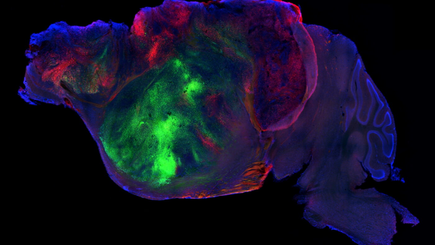
To test whether the xenocortical mice could serve as a reliable disease model, the researchers subjected the grafted animals to oxygen deprivation. Regular mice are remarkably resilient to low-oxygen conditions, but the mice with human brain cells reacted differently.
Following oxygen deprivation, the human tissue showed cellular signs of injury, and the mice developed gait problems and limb coordination difficulties.
Ethical Questions and the Boundaries of Animal Enhancement
Whenever human cells are integrated into animal models, ethical questions follow closely behind. Outside researchers have pointed out that while the technology is exceptionally powerful, deploying it in larger or longer-living animals will require careful ongoing consideration.
Sergiu Pașca, professor of psychiatry and behavioral sciences at Stanford University, emphasized that the mice do not have any enhancement, and there is no indication that what’s being created here are mice that can think like humans, or a human brain in a mouse body. Sergiu Pașca, professor of psychiatry and behavioral sciences at Stanford University
What Next for Xenocortical Research
Beyond cerebral palsy, the research team plans to use patient-derived organoids to study genetic forms of autism and frontotemporal dementia. By capturing aspects of human brain circuitry that have never before been observed in a laboratory culture—including rare nerve-cell types previously seen only in autopsied human brains—the model provides an unprecedented window into human neurobiology.
With severe psychiatric and neurodevelopmental illnesses imposing staggering medical and social burdens, researchers are eager to find out if humanized brain tissue will finally reveal the mechanisms that standard laboratory models have missed.
Worth a look

Daily Orange Juice May Alter Genes Linked to Inflammation and Blood Pressure
Drinking 500ml of pure pasteurized orange juice daily for two months changes the activity of thousands of genes...

UTHealth Houston Researchers Find Protein Signatures Predict Future Disease Risk
Researchers at UTHealth Houston discovered that cardiometabolic disease risk factors can be identified in children using protein blood...

Monaldi Hospital Study Reveals Microbiota Differences in Endocarditis Patients
Researchers in Italy have revealed notable differences in gut, oral, and skin microbiota among patients with heart valve...

Boy’s Abdominal Rash Caused by Resting Charging Laptop on Skin
A young boy presenting with a mysterious abdominal rash and suspected liver injury was ultimately diagnosed with erythema...