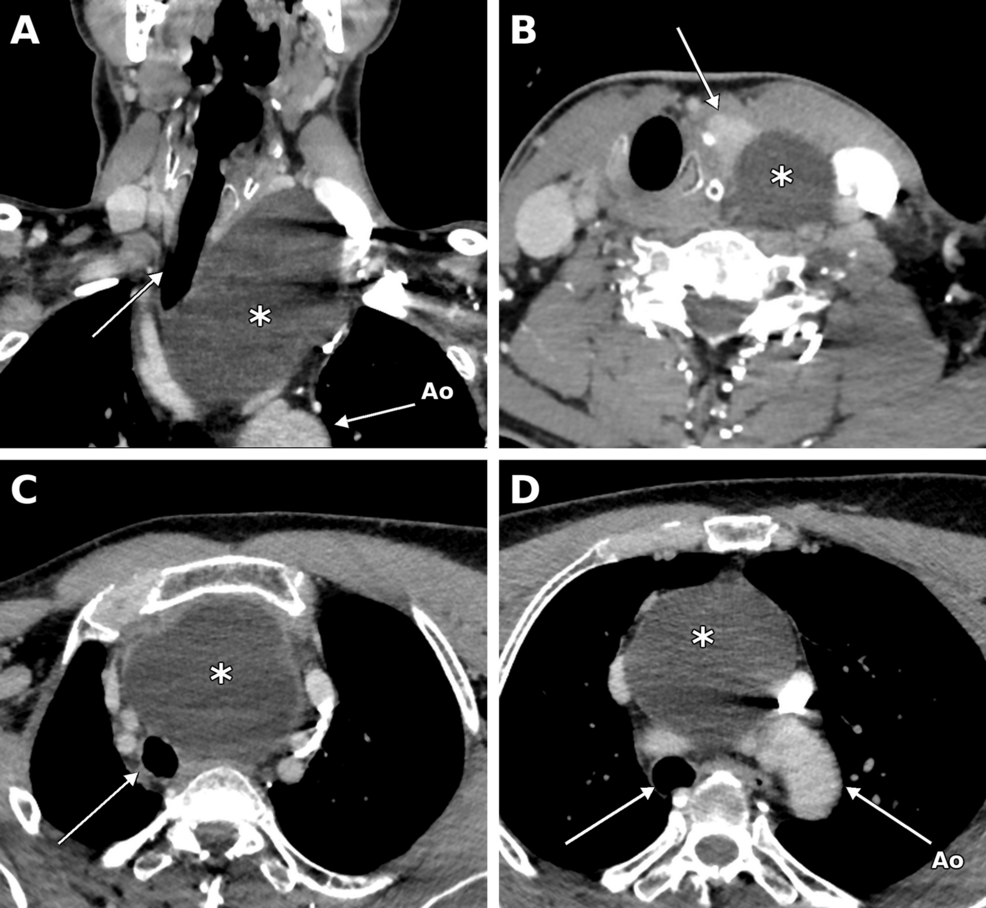
An 11-centimeter cyst, stretching from the neck down to the aortic arch, was successfully removed from a 59-year-old man in 2020, Cureus reported.
The massive lesion, which had been mistaken for a parathyroid cyst during initial diagnostic attempts, caused significant respiratory distress and left vocal fold immobility. This rare case highlights the diagnostic complexities involved when congenital anomalies present far from their typical anatomical locations.
While thyroglossal duct cysts (TGDCs) are the most frequent congenital neck anomalies, they almost always appear as midline lesions near the hyoid bone. This specific case was exceptional because the cyst extended into the anterior mediastinum, compressing the trachea and displacing the airway to the right.
The patient first experienced hoarseness and shortness of breath in February 2020. By June 2020, a computed tomography scan revealed the water-density cyst was inseparable from the left thyroid lobe and reached the level of the aortic arch.
Medical teams initially considered the lesion a non-functioning parathyroid cyst after an ultrasound-guided fine-needle aspiration yielded only clear, colorless fluid. This diagnostic uncertainty shows how difficult it is to identify deep-seated mediastinal masses that mimic other thyroid or lymphatic diseases.
Surgeons performed the excision in August 2020 using a cervical approach rather than opening the chest. To manage the high stakes of such a deep dissection, a thoracic surgeon was on standby and blood products were prepared in case a sternotomy became necessary.
The surgical team identified and protected the left recurrent laryngeal nerve before using blunt dissection to pull the 11-centimeter mass upward through the neck. The cyst was removed intact, and the patient was discharged just two days after the procedure.
Histological examination later confirmed the diagnosis. The pathology report showed an inflamed, sclerotic cyst wall associated with thyroid parenchyma, findings that are compatible with a thyroglossal duct cyst.
The rarity of this presentation is evidenced by the limited number of documented cases. A review of eight previously published mediastinal cases shows that most successful removals of such large lesions have been achieved through a cervical approach when the anatomy allows.
| Case Context | Typical Presentation | This Case |
|---|---|---|
| Primary Location | Near the hyoid bone | Extending to the aortic arch |
| Typical Size | 2.6 cm (mean) | 11 cm |
| Surgical Access | Sistrunk procedure | Transcervical excision |
The patient’s long-term outcome remains a point of clinical interest. At a follow-up in May 2025, the patient was reported to be well and had fully recovered his left vocal fold function.
Because the patient declined further CT scans, doctors could not use radiological imaging to definitively confirm the absence of recurrence. However, the clinical recovery of his vocal function serves as a primary indicator of a successful intervention.
Thyroglossal duct cysts remain a critical consideration in the differential diagnosis of cervicomediastinal cysts, even when they resemble more common parathyroid or thymic lesions.
Worth a look

Rare MYOF Genetic Variant Linked to Angioedema With Normal C1-INH Levels
A rare genetic variant in the MYOF gene may explain why a 51-year-old woman suffered from recurring angioedema...

Educators and Students Rally Against Pax Silica Initiative
Teachers, professors, and students gathered at the Bantayog ng mga Bayani in Quezon City on Thursday to voice...

Liver Proteins May Protect Brain Cells and Memory in Alzheimer’s Mice
Liver-derived proteins may offer a new way to protect brain cells and improve memory in Alzheimer’s disease models,...

Government Considering Special Session for Delimitation Bill: The Hindu
The Indian government is considering a special session of Parliament to push through the Delimitation Bill, 2026, according...