One head CT scan in childhood corresponds to roughly one extra brain tumor for every 1,266 scans performed, a massive new study published today in JAMA finds.
The research, led by UC Davis and UC San Francisco, tracked more than 3.7 million children across six U.S. health systems and Ontario, Canada, from birth for an average of 12.3 years. Among them, 1,536 brain tumors were diagnosed. The analysis, which reconstructed cumulative radiation doses to the brain from electronic medical records, showed that risk climbed steadily with higher exposure, with the strongest link seen for aggressive tumor types.
“Our findings do not mean that parents should avoid getting their children medically necessary imaging. These examinations can be lifesaving,” said Diana Miglioretti, the study’s first author and a professor and chief of the Division of Biostatistics at UC Davis Health.
“The message is that every examination using ionizing radiation should be clinically justified and performed using techniques that keep radiation dose as low as reasonably achievable while still obtaining the information needed for care.”
Brain tumors are the second most common childhood cancer and the leading cause of cancer death in children and adolescents. The study estimated that 10.1 percent of brain tumors in the cohort could be attributed to radiation from medical imaging. Exposure equivalent to a single head CT was associated with a 1.8-fold increased risk — about eight additional tumors per 10,000 children by age 21.
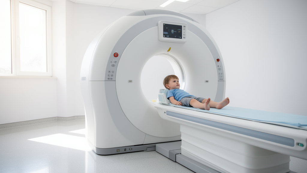
The dose-response curve
Researchers categorized cumulative brain dose and found a clear gradient. Compared with children who had no ionizing radiation exposure, those receiving more than zero but less than 1 milligray (mGy) saw a hazard ratio of 1.12. At 50 to 75 mGy — roughly the dose of a single head CT — the hazard ratio jumped to 2.30. The mean brain dose from one head CT in the study was 66.2 mGy, according to the JAMA abstract.
During follow-up, 4.3 percent of children underwent one head CT, 0.9 percent had two, and 0.4 percent had three or more. Among those diagnosed with a brain tumor, the proportions were higher: 1.6 percent had two head CTs and 1.2 percent had three or more. The association was stronger for aggressive histologies, which made up 82.5 percent of diagnosed tumors.
“Head CT is invaluable in the right setting — such as to assess for bleeding after severe trauma. However, it is overutilized, with an increasing use year after year,” said Rebecca Smith-Bindman, the study’s senior author and a professor of epidemiology and biostatistics, obstetrics, gynecology and reproductive sciences, and Health Policy Studies at UCSF.
“This study quantifies an uncommon, but serious harm resulting from radiation exposure that should be weighed every time a scan is ordered.”
Clinical justification over automatic imaging
The findings reinforce existing clinical decision rules designed to curb unnecessary scans. The Pediatric Emergency Care Applied Research Network (PECARN) rules, validated for high accuracy, help clinicians determine which children with head trauma truly need a CT.
The editorial accompanying the study in JAMA underscored the same standard: select the exam most likely to answer the clinical question, use ionizing radiation only when the expected benefit justifies the exposure, and obtain the necessary information at the lowest dose compatible with adequate image quality. The investigators stressed decisions must be driven by thorough clinical justification and active dose minimization, not automatic thresholds.
The stakes are asymmetric. A child with a minor head injury faces a tiny, delayed cancer risk from a scan that may not change management. A child with a suspected bleed faces an immediate, life-threatening risk without one. The study does not resolve that tension for any individual case — it quantifies the denominator so the conversation can start from evidence, not habit.
UC Davis Health operates inland Northern California’s only Level I trauma center for both adults and children, one of fewer than 20 such dual-verified centers nationwide. The health system traces its clinical roots to Sacramento County Hospital, established in 1852, which became a primary teaching hospital for UC Davis in 1966 and was transferred to the university in 1973.
“A small number of children will develop a brain tumor as a result of CT or other radiation tests. More judicious use of head CT and ensuring the doses for every exam are as low as possible will reduce this harm,” Smith-Bindman said.
Related reading

US Sanctions Comanchero Leaders in NZ, Australia in First for Australian Crime Group
The United States Treasury placed five men linked to the Comanchero motorcycle gang under financial sanctions on Friday,...

Latvia Parliament Approves Kulbergs Government 57-34; DBRS Flags EUR 2.9bn Fiscal Plan
Latvia's parliament approved Prime Minister Andris Kulbergs' new coalition government by a 57-to-34 vote on Tuesday evening, capping...
Microneedle Patch Tracks Troponin in Mice, Aims to Detect Silent Heart Injury After Surgery
About 300 million people undergo non-cardiac surgery every year, and most go home the same day — yet...

Teen Charged After Stealing Garda Car, Ramming Two Patrol Vehicles in Wrong-Way N25 Pursuit
A 19-year-old man has been charged with seven offences after allegedly stealing an electric Garda patrol car from...