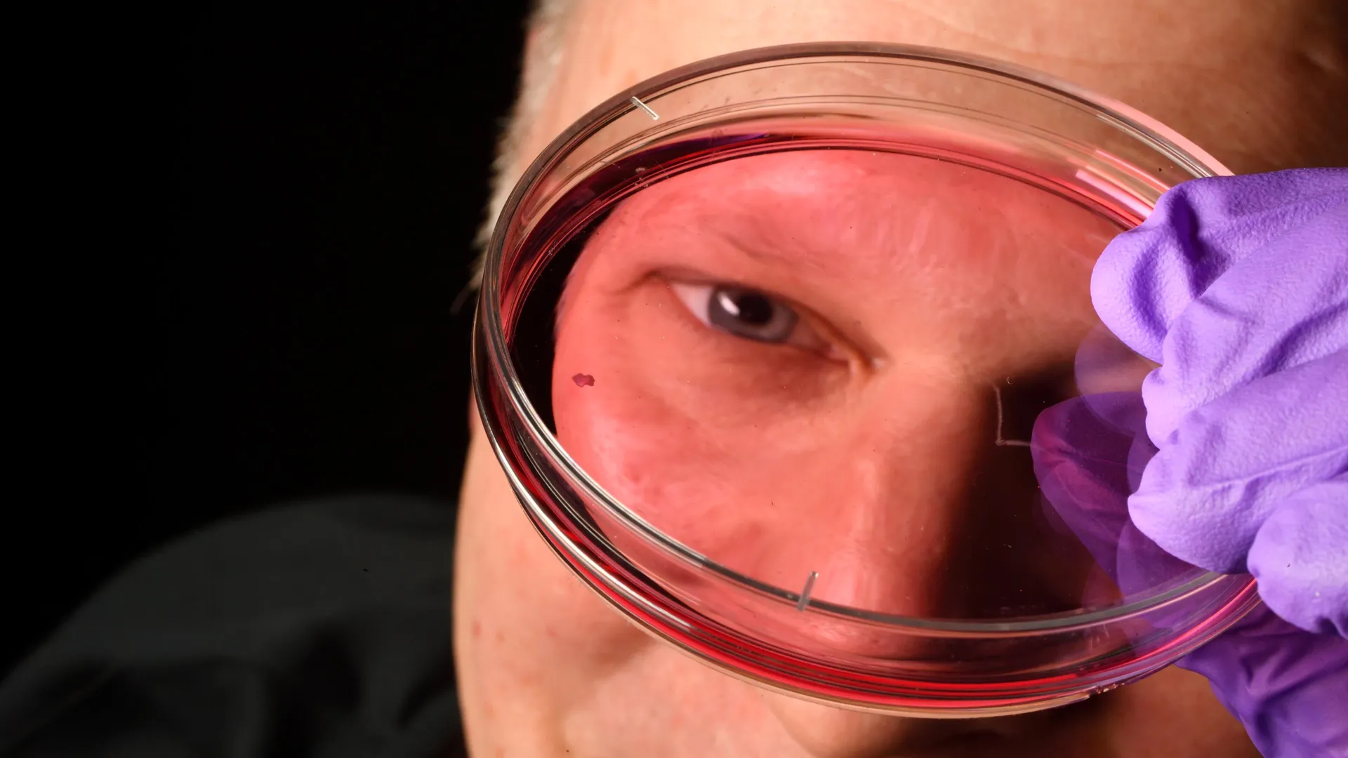
Researchers at Johns Hopkins University have identified a biological mechanism that shapes the human foveola, the retina’s center for sharp vision, by tracking how vitamin A-derived molecules and thyroid hormones convert specific cone cells during fetal development. The findings, published in the Proceedings of the National Academy of Sciences, challenge long-standing theories regarding photoreceptor migration.
Retinal Development and the Foveola Transformation
The foveola is a tiny, critical region of the retina responsible for approximately half of all human visual perception. While the rest of the retina contains a mix of blue, red, and green light-sensing cone cells, the foveola is uniquely populated by only red and green cones. For decades, the dominant scientific model proposed that blue cones formed in this central region before physically migrating outward to make room for other cell types.
New evidence from Johns Hopkins University researchers, who utilized lab-grown retinal organoids to observe fetal development, suggests a different process: the cells do not migrate. Instead, they change their identity. Between weeks 10 and 12 of fetal development, blue cones appear in the foveola. By week 14, these cells undergo a conversion process driven by two distinct mechanisms. First, retinoic acid—a molecule derived from vitamin A—is broken down to limit the creation of new blue cones. Subsequently, thyroid hormones trigger the remaining blue cones to convert into red and green cones.
“The main model in the field from about 30 years ago was that somehow the few blue cones you get in that region just move out of the way, that these cells decide what they’re going to be, and they remain this type of cell forever. We can’t really rule that out yet, but our data supports a different model. These cells actually convert over time, which is really surprising.”
Robert J. Johnston Jr., Johns Hopkins University
Structural Dynamics of RBP3 and Vision Health
Beyond the formation of cone cells, the chemical environment of the retina relies on precise molecular transport. Scientists have recently captured the native structure of Retinol-binding protein 3 (RBP3) with high accuracy, revealing it to be more dynamic than previously understood. As reported by News-Medical, RBP3 acts as a “courier” for retinoids, which are essential derivatives of vitamin A, moving them between photoreceptors and the retinal pigment epithelium.
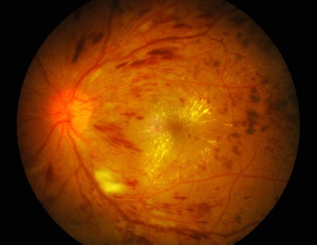
Researchers used cryo-electron microscopy to image the porcine version of this protein at a resolution of 3.67 Å. The structural analysis confirms that RBP3 contains four retinoid-binding modules that interact with various molecules, including fatty acids like docosahexaenoic acid (DHA). This protein is vital for stabilizing the retina’s biochemical environment and preventing the degradation of light-sensitive molecules. Mutations in the gene encoding RBP3 are linked to conditions such as retinitis pigmentosa, an incurable disease that leads to the gradual loss of photoreceptors.
Microgravity Risks and Future Exploration
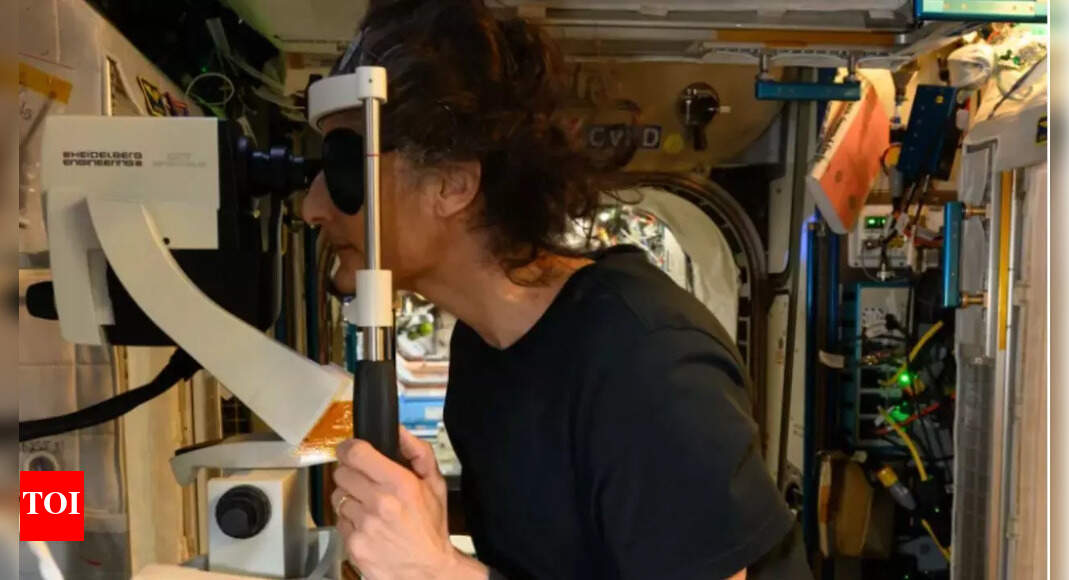
Vision challenges extend beyond biological disease to environmental stressors. NASA has documented Space-Associated Neuro-Ocular Syndrome (SANS) in astronauts during long-duration missions on the International Space Station.
Symptoms of SANS include optic disc edema and globe flattening, which can impair visual acuity. NASA continues to test countermeasures, including the “Thigh Cuff” experiment, designed to trap blood in the lower body to mitigate headward fluid shifts. Additionally, researchers are investigating the use of B vitamin supplementation and cabin environmental controls as potential stabilizers for ocular health in space.
Automation in Modern Scientific Research
The speed at which these vision-related discoveries can be translated into treatments is being influenced by the rise of self-driving laboratories. At the University of Toronto’s Acceleration Consortium, scientists are using autonomous systems to streamline drug discovery.
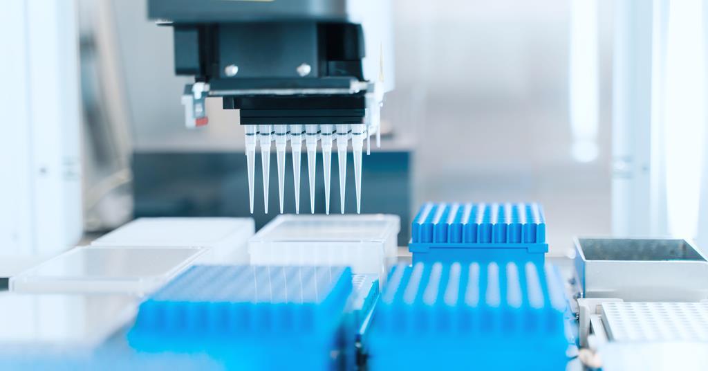
These systems utilize machine learning models that learn from each experimental cycle, allowing the lab to refine compound designs without constant human intervention. This shift toward autonomous discovery is intended to bypass the traditional bottlenecks in pharmaceutical research, where developing new therapeutics can otherwise take over a decade.
=== END ARTICLE ===
Find more reporting in our News section.
Worth a look

Quntis Monitor Light Bar Glow Features RGB LEDs and Ultrawide Support
The Quntis Monitor Light Bar Glow introduces a 20.1-inch metallic gray lighting system featuring an 88-front LED task...

Police Hunt Roommate After Waitress Jovia Namuwulya Murdered in Bukoto
Police in Kampala are investigating the murder of 22-year-old waitress and TikTok content creator Jovia Namuwulya, whose body...

US and Botswana Forces Enhance Planning During Exercise Southern Accord 2026
Exercise Southern Accord 2026 concluded on August 15 at Gaborone Technical College in Botswana, uniting military personnel from...

China Recalls Millions of Teslas Over Flush Door Handles
Minimalist electronic door latches and flush exterior handles have defined the modern electric vehicle aesthetic since Tesla popularized...