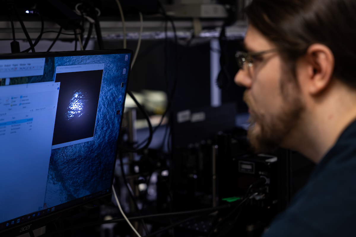
Researchers at Texas A&M University have developed an advanced imaging technology that allows for the recording of biological processes in real time at speeds of up to 1,000 frames per second. By capturing both motion and chemistry simultaneously, the team has created a method that provides a more detailed view of how biological events unfold, offering potential advancements in medical imaging and the study of disease progression. The findings, published in the *Proceedings of the National Academy of Sciences* (PNAS), detail a system that effectively eliminates the motion blur typically associated with traditional microscopic methods.
Capturing Chemistry at the Speed of Life
Traditional biological imaging tools are primarily designed to show structure, such as the shape of a cell or the outline of tissue. However, researchers emphasize that disease is fundamentally driven by chemistry—the way molecules interact, shift, and respond over time. The new technique developed by the Texas A&M team maps these chemical changes directly by utilizing the natural vibrations of molecules. According to Dr. Jizhou Wang, the paper’s lead author and a senior researcher, the team does not add anything to the system, such as dyes or labels, which can sometimes alter biological samples. Instead, they read the chemistry that is already present. The process involves using infrared light to excite molecular vibrations, which are then converted into visible light that a camera can record. Because different molecules vibrate at distinct frequencies, the system can distinguish between various chemical components within a living sample.
Eliminating Motion Blur with Picosecond Exposure
A significant challenge in live-cell imaging is motion blur, which occurs when a specimen moves during the exposure time of an image. The Texas A&M team overcame this by developing a system that captures an entire image in a single, rapid shot. Each frame is recorded on the scale of a picosecond—approximately one trillionth of a second. Physicist and co-author Dr. Alexei Sokolov, a University Distinguished Professor in the College of Arts and Sciences and associate director of the Institute for Quantum Science and Engineering, noted that at this timescale, objects do not have sufficient time to move enough to blur the image. Consequently, the resulting footage captures the system in a state very close to its natural condition. To demonstrate the technology, the team imaged living *C. elegans* worms as they moved through water. These “worm movies” preserve chemical detail while clearly showing the organisms in motion, a feat that would typically be distorted by traditional scanning methods.
Broad Implications for Biomedical Research
The ability to observe chemical processes as they happen in real time provides researchers with a more comprehensive understanding of how biological events develop moment by moment, rather than relying on a series of disconnected snapshots. “We’re able to follow processes that were essentially invisible before,” said Dr. Sokolov. “Not just where things are, but how they’re evolving, how the underlying chemistry is changing from moment to moment.” According to the researchers, this technology could have several practical applications: * Disease Tracking: Better understanding of how diseases emerge and evolve in systems where timing and chemical interactions are critical. * Therapeutic Monitoring: Detecting subtle changes in cells earlier and observing how they respond to specific treatments. * Versatility: The technique operates in water-rich environments, making it suitable for studying living organisms in their natural states. * Interdisciplinary Use: While primarily developed for biology, the team noted that the approach can be applied to other systems with rapidly changing chemistry, including physics and materials science. By overcoming the limitations of speed and blur, the Texas A&M team aims to provide a clearer window into the rapid biochemical dynamics that drive life.

Find more reporting in our Technology section.
Worth a look

Speedo Launches Speedo iQ Removable Module to Track Swimming Performance
Speedo is making its entry into the wearable technology market with a compact triangular module designed to fit...

Windscribe Offers Pro VPN Subscription With Permanent Price Lock Deal
Windscribe is offering its Pro subscription for thirty-nine dollars for a full year, representing a sixty-four percent discount...
Chevron Signs 20-Year Power Agreement to Supply Microsoft Data Center
Chevron signed a 20-year power purchase agreement with Microsoft to supply electricity from natural gas turbines to a...

Xiaomi 18 Fold Leaks Reveal Wider Design and XRING O3 Chip
Xiaomi has confirmed a September 2026 launch in China for its newly branded 18 Fold, moving the device...