
Scientists Reveal Kinesin-1 Activation Mechanism
Researchers have uncovered the structural mechanics behind how the motor protein kinesin-1 is activated within nerve cells. The findings, published July 15 in Science Advances, provide a detailed look at how cells regulate this protein to transport essential cargo, such as neurotransmitters, along microtubule tracks.
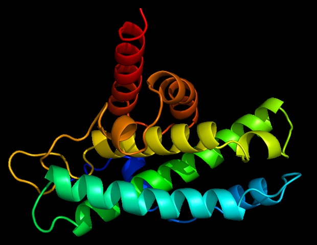
Kinesin-1 has long been compared to a robotic broomstick that travels at 100 steps per second. While its movement mechanism—powered by ATP—has been understood for decades, the method the cell uses to turn the protein on and off remained a mystery. By using cryo-electron microscopy to image the protein in its inactive state, a team led by researchers at the University of California, Davis, discovered that the inactive kinesin-1 is folded in half. In this state, the protein’s top end is wedged between its legs, and a connector acts as a “rubber band” to lock the structure, preventing movement and obstructing the cargo-attachment site.
The study found that the cell activates the protein by utilizing a partner protein called MAP7. When MAP7 attaches to kinesin-1, it wedges into the structure and pops the rubber band loose. This action causes the protein to unfold, freeing its legs and exposing the docking site for cargo transport.
Implications for Neurodegenerative Disease
The ability to visualize this “on-off” switch is significant because kinesin-1 malfunctions are linked to several incurable neurodegenerative diseases, including amyotrophic lateral sclerosis (ALS), Charcot-Marie-Tooth Disease Type 2, and hereditary spastic paraplegia 10. These conditions often arise from mutations that prevent the protein from correctly transitioning between its active and inactive states.
“If you want to design drugs, having these clear structures will be a major advance for that,” said coauthor Richard McKenney, a professor of molecular and cellular biology at UC Davis. Lead author Jawdat Al-Bassam added that the discovery could lead to the design of molecules that bind to mutant proteins to correct their defects. The research was funded by the National Institutes of Health and involved collaboration with researchers from Johns Hopkins University.
Find more reporting in our Science section.
Related reading
Astronomers Discover 10-Sided Atmospheric Wave at Saturn’s South Pole
Astronomers using the Hubble Space Telescope have discovered a massive, evolving 10-sided atmospheric wave circling Saturn’s south pole.
Monash Researchers Propose Expanding Genus Homo to Rewrite Family Tree
Monash University researchers argue that early human relatives and close fossil relatives living up to 5 million years...
Researchers Measure Gravity Effect on Quantum Atoms in New Study
Researchers have directly measured gravity’s predicted effect on the quantum phase of freely falling rubidium atoms, observing Einstein’s...

Corrected Headline: Harvest Moon Rises Near Saturn on September 26
The Harvest Moon of September 2026 will rise on September 26, appearing near Saturn and Neptune in the...