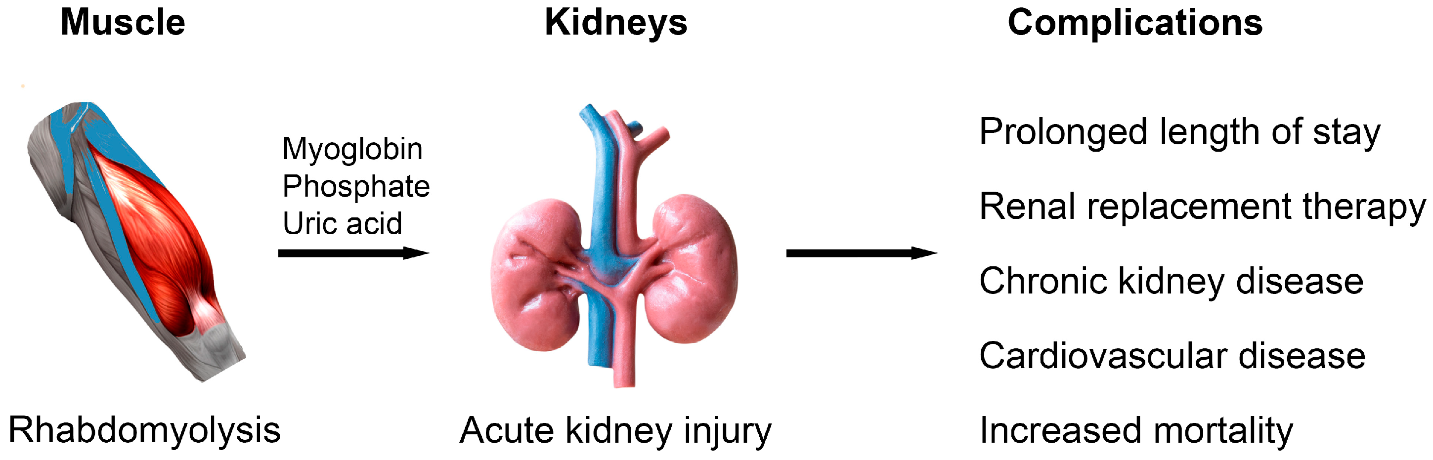
Massive amphetamine-associated rhabdomyolysis can trigger severe metabolic, cardiovascular, and renal complications, including acute kidney injury requiring dialysis, as well as complications such as muscular calcinosis, according to clinical case documentation.
Severe muscle breakdown can release massive quantities of intracellular components into the bloodstream, posing profound risks to renal and cardiac function. While rhabdomyolysis is broadly understood as a clinical syndrome involving skeletal muscle injury, specific toxic exposures can drive creatine kinase concentrations to near-million levels, testing the limits of intensive hospital management.
Case Findings and Dialysis-Requiring Kidney Injury in Two Patients
Recent medical documentation detailed two cases of severe amphetamine-associated rhabdomyolysis complicated by acute kidney injury requiring renal replacement therapy. The first patient, a 46-year-old man, presented with an exceptionally high serum creatine kinase level of 947,699 U/L, a serum creatinine of 734 µmol/L, and hyperkalemia measuring 6.8 mmol/L.
Medical teams administered supportive care alongside 11 sessions of intermittent hemodialysis. Excessive sympathetic stimulation from amphetamine intoxication drives hyperthermia, psychomotor agitation, increased muscular activity, peripheral vasoconstriction, dehydration, and direct myocyte toxicity. These converging mechanisms cause massive skeletal muscle necrosis and the release of myoglobin, creatine kinase, potassium, and phosphate into circulation.
Pathophysiology of Myoglobin-Mediated Renal Damage
When muscle tissue breaks down on a massive scale, circulating myoglobin frequently leads to pigment-induced acute kidney injury.
Renal injury develops through several distinct pathways, including intrarenal vasoconstriction, oxidative tubular injury, direct tubular toxicity, and intratubular cast formation. Best predictors for acute kidney injury development in these clinical scenarios include volume depletion, elevated initial serum creatinine, low serum bicarbonate, hypocalcemia, and hyperphosphatemia. Additional factors like hypoalbuminemia and increased blood urea nitrogen also associate with renal decline.
Muscular Calcifications and Delayed Hypercalcemia Observed
Beyond immediate renal failure, massive rhabdomyolysis can provoke unusual and severe secondary developments. The 46-year-old patient experienced transient left ventricular systolic dysfunction, eventually recovering normal left ventricular ejection fraction. However, on hospital day 28, the patient developed severe corrected hypercalcemia measuring 4.10 mmol/L following the profound hypocalcemia observed during the initial phase of muscle breakdown.
Computed tomography imaging and etiological evaluations revealed extensive pathology. Imaging revealed diffuse muscular calcifications consistent with metastatic muscular calcinosis secondary to severe rhabdomyolysis. The clinical course ultimately concluded with cardiac arrest and death, though investigators noted that a direct causal relationship between the delayed hypercalcemia, transient systolic dysfunction, and the fatal cardiac arrest could not be definitively confirmed alongside possible amphetamine-associated cardiotoxicity.
Broader Medical Context of Non-Traumatic Muscle Breakdown
Medical literature distinguishes between traumatic causes—such as crush injuries, prolonged immobilization, and natural disasters—and non-traumatic etiologies. Non-traumatic categories encompass viral illnesses, metabolic myopathies, seizures, inherited disorders, extreme exercise, alcohol abuse, toxins, and medications. Among pharmaceuticals, statins are recognized as the most common class to cause muscle pain and occasionally rhabdomyolysis, while specific agents like propofol can trigger propofol infusion syndrome, featuring metabolic acidosis and rhabdomyolysis.
Diagnosis typically relies on marked elevations in serum creatine kinase, though no single established diagnostic serum level universally applies. Clinicians frequently utilize thresholds ranging from 3 to 5 times the upper limit of normal up to clinical definitions requiring eleven times normal alongside pigment-induced nephropathy. Management centers on active airway, breathing, and circulation assessment, appropriate hydration to maintain end-organ perfusion, close urine output monitoring, electrolyte correction, and the prompt identification of complications like compartment syndrome and disseminated intravascular coagulation.
Related reading

Researchers at the University of Virginia developed new imaging technique to study Alzheimer’s disease metabolism and oxygen use
Researchers at the University of Virginia combined two-photon fluorescence lifetime imaging microscopy with phosphorescence lifetime imaging microscopy in...
Marjan Monfared Develops PrevenTherm System to Detect Pressure Injuries
Marjan Monfared, a fellow of the 2026 Women Who Empower Innovator Awards, has developed the PrevenTherm Monitoring System...

Researchers at the University of Washington’s David Baker lab develop working molecular scissors
Researchers at the University of Washington Institute for Protein Design have used artificial intelligence to engineer synthetic proteases...
Young Adults’ Stroke Risk Nearly Doubles Over Three Decades
Medical data tracking Greater Cincinnati and Northern Kentucky over nearly three decades reveals stroke rates among adults aged...