
Retinal displacement after detachment repair shows up in roughly one in eight eyes when surgeons use the most common modern technique, while an older method keeps that rate near zero, the latest case series found.
The gap — 15.5% after pars plana vitrectomy versus 2.2% after scleral buckle — comes from a single-center review of 194 eyes published in Ophthalmic Surgery, Lasers & Imaging Retina last October. Researchers at the same institution also reported a 17.6% displacement rate when the two procedures were combined. “Retinal displacement after RRD repair is more prevalent after PPV,” the authors concluded, noting that visual acuity did not differ significantly between eyes with and without the shift.
That finding lands in the middle of a broader debate. A narrative review in Cureus this year surveyed the last decade of literature and found reported displacement rates swinging wildly from 7% to 62.8% depending on the imaging platform and surgical approach. An earlier systematic review on PMC, whose search closed in August 2022, confirmed the same spread across 22 studies and flagged facedown positioning, perfluorocarbon use, and proliferative vitreoretinopathy as recurring risk factors.
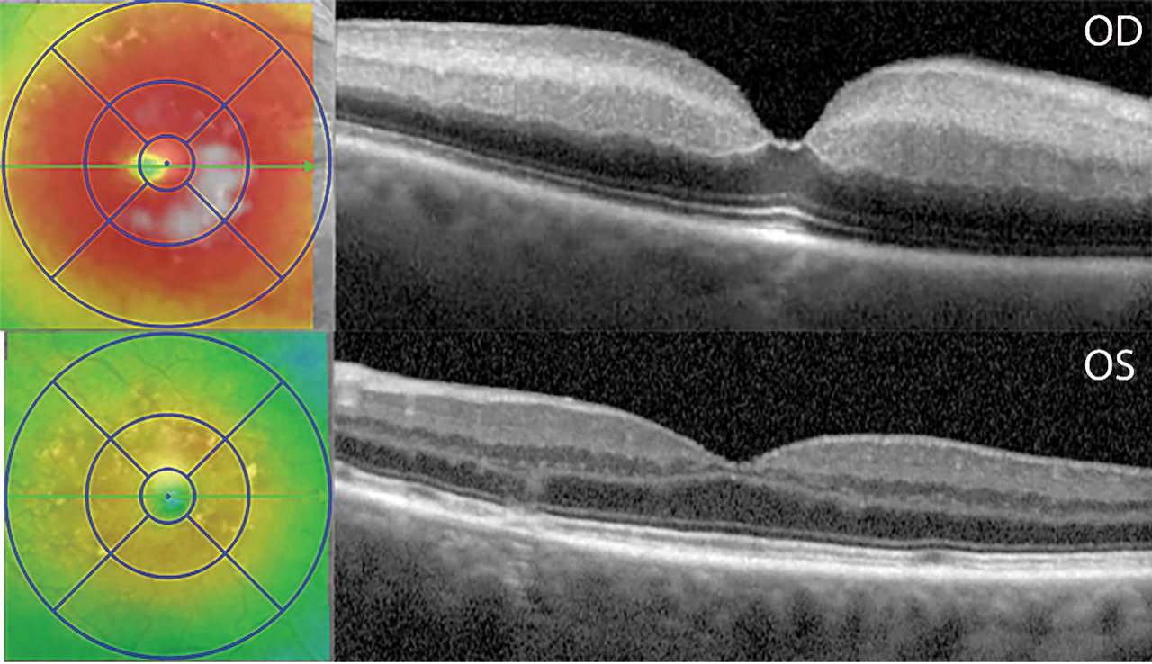
What the patient actually sees is the unresolved question. The Cureus review highlighted a prospective cohort where 10 of 17 patients with documented retinal shift reported metamorphopsia — distorted vision — yet their visual acuity scores stayed statistically normal. Another study graded displacement on fundus autofluorescence: grade 1 showed an attached shadow, grade 2 a clear separation, and grade 3 a shift exceeding two vessel diameters. Only the higher grades correlated with worse final acuity.
Detection itself is part of the problem. Fundus autofluorescence, the standard screening tool, missed nearly half the cases that infrared homography caught in one comparative study — 43.7% versus 93%. Blue and green ultra-widefield autofluorescence performed similarly, around 45% detection overall, while confocal scanning laser ophthalmoscopy and fundus-camera FAF agreed only moderately. Optical coherence tomography angiography adds another layer, linking foveal avascular zone circularity to both aniseikonia scores and maximum displacement amplitude.
“Retinal displacement adds another layer to this evaluation. It may help explain why some patients continue to experience visual symptoms despite successful reattachment,” the Cureus authors wrote.
Microstructural changes on OCT tell a parallel story. Eyes without detectable displacement — labeled high-integrity retinal attachment — show photoreceptors returning close to their original position relative to the retinal pigment epithelium. Eyes with significant vessel printings, termed low-integrity attachment, more often display ellipsoid zone disruption, external limiting membrane breaks, and retinal folds, all associated with poorer visual quality even when the retina is technically flat.
The 2024 case series, conducted between April 2020 and February 2022, used Optos ultra-widefield autofluorescence and found no acuity penalty for displacement at the group level. But the ALIGN study, cited in the Cureus review, tied pars plana vitrectomy to worse aniseikonia scores and worse best-corrected visual acuity compared with pneumatic retinopexy, linking the displacement itself to the aniseikonia rather than making it the sole explanation for the acuity drop.
Surgeons still lack a unified grading system, a consensus on which imaging modality to trust, and prospective data connecting specific displacement magnitudes to patient-reported outcomes. The next step, several reviews argue, is standardizing detection — probably with multimodal imaging — and tracking whether surgical modifications, such as avoiding perfluorocarbon or altering tamponade strategy, can shrink the gap between anatomical success and the vision patients actually live with.
Worth a look

Bengaluru Man Kills Stepson, Locks Wife in Room for Four Days; Arrested
Bloodstains on the floor and a woman screaming from behind a locked bedroom door greeted the landlord and...

BNPB Places 12 Provinces on Heavy-Rain Alert, Warns of Floods and Landslides
The National Disaster Mitigation Agency placed 12 Indonesian provinces on heavy-rain alert Friday, warning that flooding and landslides...

Daraxonrasib Plus Chemo Halts Tumours in 90% of Pancreatic Cancer Patients, Trial Shows
In a trial of 61 people with advanced pancreatic cancer, tumours stopped growing in 90 percent of them...

School Bus Fees to Jump 250% to €350 Next September Under Budget 2027
The annual school bus ticket for a secondary student will jump from €100 to €350 next September, a...