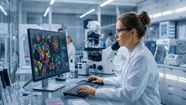
Cellpose and StarDist, two open-source tools, are revolutionizing deep learning cell segmentation in biological imaging, with distinct approaches tailored to different imaging challenges. According to Technology Networks, researchers rely on these tools to automate a task once done manually, but choosing between them depends on cell shape and imaging conditions. The broader shift toward AI-driven analysis reflects a trend reshaping biological research.
Automation of Cell Segmentation
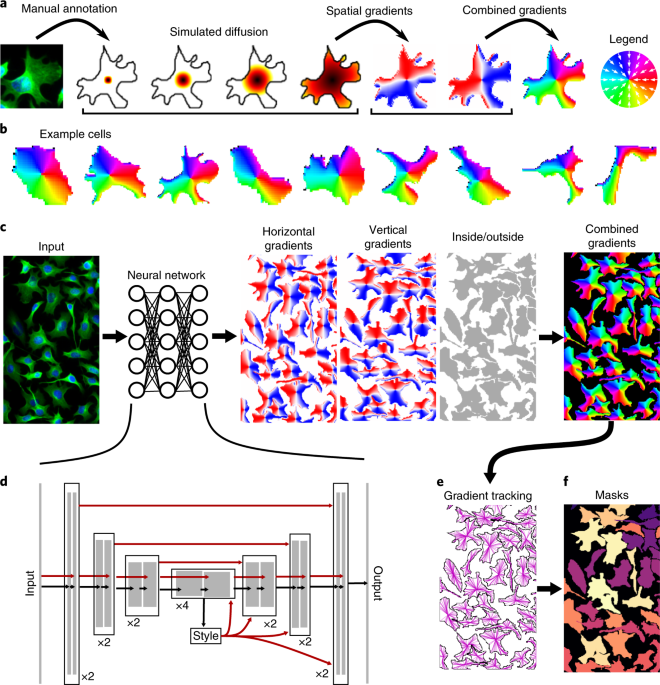
Technology Networks reported that Cellpose and StarDist have become essential for automating cell segmentation, a process critical for biological analysis. Manual methods are impractical for large datasets, and traditional algorithms struggle with overlapping cells or varying shapes. Deep learning tools like Cellpose and StarDist address these issues by learning from diverse datasets rather than fixed rules. “Cellpose predicts a generalist flow representation that works across a wide range of cell shapes and imaging modalities without retraining,” the article noted. StarDist, on the other hand, excels in densely packed, round nuclei by modeling them as star-convex polygons, a method that avoids merging touching cells.
Choosing the Right Tool
The choice between the two tools hinges on specific imaging needs. Cellpose’s versatility makes it suitable for varied cell types and imaging conditions, while StarDist’s precision is ideal for crowded, round nuclei. “Choosing between the two depends primarily on cell or nucleus shape, packing density, and imaging modality rather than on any single accuracy benchmark,” Technology Networks wrote. This distinction is crucial for labs handling diverse experiments, as a tool optimized for one scenario may fail in another.
Broader Transformation in Imaging
Beyond their technical differences, the tools reflect a broader transformation in biological imaging. Classical methods required extensive parameter tuning for each new experiment, limiting scalability. Deep learning approaches eliminate this burden, enabling researchers to focus on analysis rather than setup. “The shift toward algorithmic, data-driven analysis they represent sits within a broader set of biological imaging analysis tasks that AI has transformed over the past several years,” the article stated. This trend underscores AI’s growing role in accelerating scientific discovery.
Open-Source Collaboration
The development of these tools also highlights the importance of open-source collaboration. Cellpose 3, for instance, includes one-click image restoration for noisy data, a feature that streamlines workflows. Meanwhile, newer models adapted from general-purpose foundation models suggest the field is moving toward even more adaptable solutions. “Newer segmentation models adapted from general-purpose foundation models point to where the field is heading next,” Technology Networks noted. This evolution could further reduce the need for custom training, making advanced analysis accessible to a wider range of researchers.
While the tools offer significant benefits, their effectiveness relies on proper application. Errors in segmentation can propagate through downstream analyses, such as cell counting or feature extraction, leading to unreliable results. Every downstream measurement… inherits whatever errors the segmentation step introduced, the article warned. Researchers must therefore carefully evaluate which tool aligns with their specific imaging challenges.
The broader implications of these advancements extend beyond individual labs. As AI continues to refine biological imaging, it could enable large-scale studies that were previously infeasible. For example, high-throughput screening of drug candidates or detailed analysis of cellular dynamics in complex tissues may become more routine. “The stakes of getting segmentation right extend well beyond the time saved on any single image,” the article emphasized.
The collaborative nature of these tools also fosters innovation. By building on shared frameworks, researchers can build upon each other’s work, accelerating progress. “Each tool approaches the underlying prediction problem differently enough that neither is a strict replacement for the other,” Technology Networks wrote. This diversity of approaches ensures that the field remains adaptable, with multiple solutions available for different needs.
Looking ahead, the integration of these tools with emerging technologies could further enhance their capabilities. For instance, combining Cellpose or StarDist with advanced imaging techniques like super-resolution microscopy might unlock new insights into cellular structures. “The field is heading next” toward even more sophisticated models, as the article suggested. However, the success of these innovations will depend on continued research and open collaboration.
For researchers, the key takeaway is to match their tools to their specific challenges. While Cellpose offers broad applicability, StarDist’s specialized design may be indispensable for certain experiments. As the field evolves, staying informed about these tools’ strengths and limitations will be critical. “Understanding both remains useful even for labs that ultimately settle on one as a default,” the article concluded.
The impact of these tools on biological research is already evident, but their full potential may only be realized through ongoing refinement and adaptation. As AI continues to shape scientific workflows, the distinction between manual and automated analysis will blur, opening new avenues for discovery.
Find more reporting in our Science section.
Related reading

Moon to Occult Jupiter Over Eastern North America on October 6
Skywatchers across much of eastern North America can watch a rare lunar occultation early Tuesday morning as a...

Saturn Reaches Closest Approach to Earth for the Year
Saturn stands roughly 784 million miles from Earth as opposition positions the ringed giant at its closest annual...

Researchers Identify Candidate Second-Generation Planet Orbiting White Dwarf
Circling its parent dead star every 4.4 days, a Jupiter-sized gas giant orbits the white dwarf HS 0209+0832...
UK Proposes Norfolk Housing Levy to Unlock Stalled Homes
Environment Secretary Angela Eagle launched a public consultation on 17 September for a Norfolk pilot scheme requiring housing...