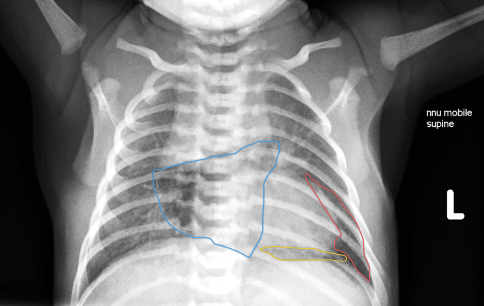
When a routine neonatal check turns up a rare triple air leak without sending an infant into acute cardiac arrest, doctors gain a fascinating window into how resilient—and unpredictable—newborn physiology can be. In a recent case report published in the medical journal Cureus, clinicians detailed the unexpected diagnosis of a term neonate who developed concurrent pneumothorax, pneumomediastinum, and pneumopericardium following brief delivery-room respiratory support.
A Routine Delivery Followed by Persistent Tachypnoea
The infant, born at 39 weeks and two days gestation by a category 2 caesarean section due to failure to progress, arrived into a high-vigilance clinical environment. The mother’s medical history included gestational diabetes mellitus managed with metformin and hypothyroidism treated with levothyroxine. More critically, her membranes had ruptured 34 hours prior to delivery, and she developed an intrapartum pyrexia of 37.8°C, receiving intravenous antibiotics just before the birth.
The baby boy weighed 3.46 kg and scored strong Apgar ratings of 9, 9, and 10 at one, five, and ten minutes. Yet, at three minutes of life, cyanosis set in, dragging oxygen saturation down to 67%. The neonatal team swiftly applied positive end-expiratory pressure (PEEP) at 5 cmH2O with an initial fraction of inspired oxygen of 21%, which was later up-titrated to 80%. When PEEP was withdrawn around the seven-minute mark, his saturation dipped again, prompting continued support for one hour until he could breathe comfortably in ambient air.
Initial medical consensus attributed the early respiratory compromise to transient tachypnoea of the newborn (TTN) or a delayed cardiorespiratory transition. But the tachypnoea refused to fade. Over the subsequent days, his respiratory rates hovered stubbornly between 44 and 79 breaths per minute on day one, settling into a persistent 50 to 68 breaths per minute through days two and four. Despite the rapid breathing, his heart rate stayed normal at 118 to 142 beats per minute, his temperature remained stable, and he fed adequately, dropping a modest and expected 3.6% in body weight.
The Day 4 Radiograph That Changed the Picture
Because the mother presented with a prolonged rupture of membranes and fever, the clinical team quite naturally suspected early neonatal sepsis. On day three, they initiated a partial septic screen. C-reactive protein registered at a reassuringly low 4.6 mg/L, and blood cultures showed no growth at 36 hours. But with the tachypnoea lingering into day four, physicians ordered a repeat blood gas, a fresh CRP check, a precautionary 48-hour course of empirical intravenous antibiotics, and a chest radiograph.
That single x-ray revealed a dramatic trifecta: a left anterior pneumothorax, pneumomediastinum, and pneumopericardium, all sitting quietly side by side without any accompanying lobar collapse or consolidation. Pneumopericardium remains the rarest of these three pulmonary air leaks, carrying a distinct clinical hazard because even a modest volume of air trapped in the pericardial space can trigger cardiac tamponade. Yet, this infant displayed no hemodynamic instability.
Managing the case called for careful observation rather than surgical intervention. Doctors opted for conservative treatment, relying on close cardiorespiratory monitoring and serial imaging. A follow-up x-ray performed 48 hours later confirmed that both the pneumothorax and pneumopericardium had completely resolved, leaving only a stable, shrinking pneumomediastinum. The infant finished his 48-hour antibiotic course as blood cultures returned clean and CRP fell to 1.5 mg/L, and he was discharged home.
What This Rare Presentation Means for Neonatal Care
By day 12 of life, an outpatient review confirmed that the pneumomediastinum had completely vanished, leaving the child with no lingering health concerns. The case serves as a striking reminder that multi-compartment air leak syndromes do not always announce themselves with sudden, catastrophic decompensation. Sometimes, they hide behind the subtle, persistent rapid breathing that clinicians might easily misattribute entirely to infection or delayed lung fluid clearance.
While delivery-room PEEP provides a plausible mechanical trigger via barotrauma and alveolar overdistension—where air dissects along perivascular sheaths into the mediastinum and pleural or pericardial spaces—spontaneous air leaks remain a possibility even after minimal pressure support. Ultimately, the Cureus report highlights the clinical value of maintaining a low threshold for chest radiography when evaluating neonates with unexplained, ongoing respiratory findings.
Keep reading

Drinking tea may help protect your heart, but there’s a surprising catch – Science Daily
How a Massive Nine-Year Study Links Daily Tea Consumption to Vascular Health Pouring a warm cup of tea...

Four people died after entering a waste chamber at a private hotel in Ujjain late Saturday
Four people died after entering a waste chamber at a private hotel in Ujjain late Saturday, according to...

Google Project Suncatcher AI Generates First Haiku From Orbit
Google successfully operated an artificial intelligence model aboard a prototype satellite in orbit, receiving a computer-generated haiku back...

Empowering Women as Knowledge Creators through Open Education Initiatives
When Tri Astari addressed the Open Education Conference 2026 (OpenEd26) on October 27, 2026, she highlighted that increasing...