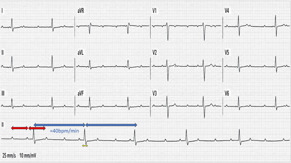
Complete atrioventricular block shows up at diagnosis in only 2.8% of infective endocarditis patients, according to the 2023 European Society of Cardiology guidelines — yet when it appears, it is one of the most ominous signals in cardiology, because the infection has usually already reached the heart’s electrical wiring. Cureus reported just such a case: a previously healthy 25-year-old woman who arrived at an emergency department with a two-day history of profound weakness, a heart rate of 40 beats per minute, and a complete third-degree AV block on her first ECG — and who, despite a blood pressure that initially read a deceptively normal 110/80, was already in the grip of one of the most destructive cardiac infections documented.
The aortic root sits directly against the atrioventricular node and the bundle of His, which means an abscess forming around the aortic valve can physically invade the conduction system. That is why new-onset AV block in a patient with fever and a new murmur is treated as a red flag for periannular extension, and why transesophageal echocardiography is rushed into play. In this woman’s case, the imaging told a devastating story.
A 45 × 27 mm Abscess, Three Valves Under Attack, and a Heart Still Pumping Normally
Urgent transthoracic and transesophageal echocardiography revealed extensive destruction: a tricuspid aortic valve with a large, highly mobile 20 × 12 mm vegetation involving the right coronary and noncoronary cusps, flail of the right coronary cusp, and severe aortic regurgitation confirmed by a broad turbulent jet and holodiastolic flow reversal in the descending aorta. A periannular cavity measuring 12 × 9 mm with color Doppler flow suggested a communicating pseudoaneurysm, and TEE went further, identifying a large aortic root abscess of roughly 45 × 27 mm extending toward the interatrial septum — which itself was markedly thickened at its base. A 13 × 9 mm vegetation sat on the mitral valve with moderate mitral regurgitation, and an 18 × 10 mm vegetation protruded into the right atrium. Tricuspid regurgitation rounded out the multivalvular involvement.
Here is the detail that makes the case so striking: her left ventricle was non-dilated, with an end-diastolic diameter of 53 mm and preserved systolic function. The acute nature of the severe aortic regurgitation was supported by the absence of ventricular dilatation and a non-widened pulse pressure of 30 mmHg. The failure was electrical and hemodynamic — cold extremities and a four-second capillary refill signaled peripheral hypoperfusion — and within hours the patient progressed to cardiogenic shock and fatal cardiac arrest.
Why This Case Differs From the Textbook Survival Story
The same complication — complete AV block from endocarditis — appears in a second report indexed on the U.S. National Library of Medicine’s PubMed Central, and the contrast between the two patients is instructive. There, a 38-year-old man with a bicuspid aortic valve developed complete AV block four days into antibiotic treatment for Staphylococcus aureus endocarditis after an orthodontic procedure; a temporary pacemaker was placed, a fistula around the AV node was found at surgery, the aortic valve was replaced the next day, and he recovered with no subsequent cardiac events. The Cureus patient, by contrast, had an unknown pneumococcal vaccination status, no identifiable portal of entry, no classic skin findings, and fulminant Streptococcus pneumoniae infection that outran intervention.
What happens next in cases like this is already written into the guidelines: urgent surgical management when periannular extension produces conduction disturbance, with pacing equipment immediately available during imaging. The woman in the Cureus report had that equipment present during her echocardiograms, and still the infection had already destroyed too much tissue. The electrical instability — a slow, narrow-QRS escape rhythm of about 40 beats per minute with complete atrioventricular dissociation — heralded the catastrophic collapse rather than merely accompanying it.
For anyone who has ever dismissed a fever as minor, this case is the counterargument: two weeks earlier she had a self-reported temperature of 38°C, and two days before admission she felt profound asthenia. By the time the ECG showed the block, the abscess had already crossed into the interatrial septum. The heart’s electrical system gave the alarm. The question the case leaves open is whether anything earlier — vaccination, earlier recognition of that first fever — would have changed the trajectory, since her pneumococcal vaccination status was never established.

WHO authorizes Ebola vaccine trials for DRC Bundibugyo outbreak
The World Health Organization has authorized clinical trials for two new Ebola vaccines, ChAdOx1 BDBV and mRNA-1469, to...

South Africa to be suspended from AGOA starting January 2027
Washington Cuts South Africa Loose From AGOA as the Rand Holds Steady The rand barely flinched when Washington...

High Court to Hear Pauline Hanson’s Appeal Over Racial Discrimination Ruling
High Court Agrees to Hear Pauline Hanson's Racial Discrimination Appeal The High Court of Australia has consented to...

Gene Therapy Reduces Seizures by 90% in Epilepsy Clinical Trial
Three adults whose epilepsy had resisted at least two anti-seizure medications saw their monthly seizures fall by more...