A 27-year-old woman nearly died from a life-threatening complication of a sore throat after an initial period of clinical improvement masked the progression of a rare infection, Cureus reported.
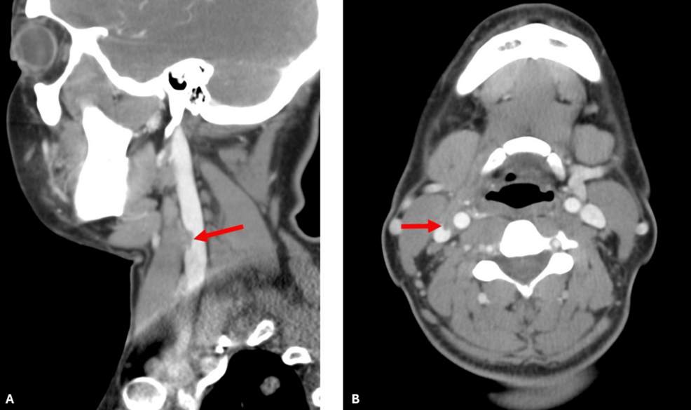
The patient developed Lemierre’s syndrome, a condition where an oropharyngeal infection spreads to the internal jugular vein. This progression leads to septic thrombophlebitis, bacteremia, and the movement of septic emboli, which frequently settle in the lungs.
While the syndrome was first described by André Lemierre in 1936, it has seen a resurgence in recent years. This shift is largely attributed to better diagnostic imaging and a higher clinical suspicion among medical professionals, particularly in adolescents and young adults.
Most cases of Lemierre’s syndrome are driven by Fusobacterium necrophorum, an anaerobic organism that triggers tissue necrosis and thrombosis. However, this specific case involved Streptococcus constellatus, an uncommon pathogen for this syndrome.
The patient’s journey began with symptoms that appeared to be a standard, albeit severe, illness. She presented to the emergency department with two days of nausea, vomiting, and diarrhea, alongside a 103°F fever, sore throat, and chills.
Initial evaluations showed significant distress. Her heart rate reached 140 beats per minute, and her blood pressure was a low 104/66 mmHg. Physical exams revealed swelling on the right side of her neck and bilateral tonsillar swelling with purulence.
After receiving intensive treatment, including intravenous ceftriaxone, piperacillin/tazobactam, and vancomycin, the patient showed signs of recovery. By hospital day seven, her symptoms had largely resolved, and she was discharged on oral ampicillin and acetaminophen.
This temporary improvement proved to be a dangerous diagnostic trap.
Just one day after returning home, the patient was readmitted with new-onset abdominal and flank pain. While her initial symptoms seemed to have vanished, the infection had moved deeper into her system.
The second hospital admission revealed the true scale of the infection. A CT scan of the chest identified multiple new bilateral pulmonary nodules, several of which were cavitary, suggesting the presence of septic emboli.
Further imaging through duplex ultrasonography of the neck confirmed the diagnosis of Lemierre’s syndrome by detecting thrombosis in the right internal jugular vein. The presence of these nodules in the context of a recent throat infection is a hallmark of the condition.
The clinical data from her second admission highlighted the systemic impact of the infection:
| Clinical Finding | Observation |
|---|---|
| Pulmonary Status | Cavitary pulmonary nodules (septic emboli) |
| Vascular Status | Right internal jugular vein thrombosis |
| Secondary Infection | Sepsis secondary to acute pyelonephritis |
Because the infection was polymicrobial and involved S. constellatus, the risks were heightened. Research indicates that polymicrobial infections in these cases can lead to an elevated 30-day mortality rate compared to monomicrobial infections.
Following the diagnosis, medical teams moved from broad-spectrum antibiotics to a targeted regimen. The patient was prescribed a six-week course of outpatient intravenous ceftriaxone and metronidazole, along with systemic anticoagulation using apixaban.
The patient eventually recovered without the need for surgical intervention. The case shows that in oropharyngeal infections, a sudden period of feeling better does not always mean the danger has passed.
Related reading
Six Family Members Killed in Massive Fire at Goldsmith’s House in Madhya Pradesh’s Seoni
Six people died in a massive fire that engulfed a three-storey building in Madhya Pradesh, turning a festive...

Cervical Cancer Awareness and Screening Attitudes Among Women: A Cross-Sectional Study in Bangladesh
Awareness gaps persist in Dhaka cervical cancer screening Most women attending a major hospital in Dhaka, Bangladesh, have...

Pedestrian Dies Following M20 Motorway Collision in Limerick
Pedestrian dies in M20 motorway collision A pedestrian has died after being struck by a car on the...

Typhoon Cempaka to Enter PAR Monday; No Landfall Expected
Typhoon Cempaka approaches Philippine waters Typhoon Cempaka is expected to enter the Philippine Area of Responsibility (PAR) on...