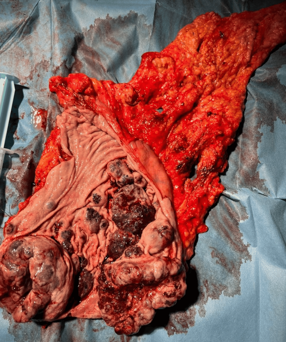
A 73-year-old man walked into an emergency room exhibiting severe gastrointestinal bleeding, yielding a remarkably rare medical case study involving primary gastric melanoma that tested negative for the key SOX10 marker, Cureus reported.
The patient arrived with symptoms of hematemesis, melena, abdominal pain, and fatigue. Initial medical examinations revealed acute-on-chronic blood loss anemia alongside tachycardia and epigastric tenderness. Endoscopy subsequently uncovered irregular, dark, multifocal submucosal lesions spread across the gastric antrum, corpus, and fundus. Because initial biopsies remained inconclusive, a computed tomography (CT) scan was ordered, revealing an irregular parietal lesion that forced an emergency total gastrectomy due to acute gastric hemorrhage.
Macroscopic and microscopic analysis of the resected tissue displayed multilocular submucosal pigmented lesions, with vascular and neural infiltration extending entirely through the stomach wall into the periventricular adipose tissue. Out of 15 regional lymph nodes examined, five showed infiltration by epithelioid pigmented cells.
Immunohistochemical Anomalies and Diagnostic Hurdles
The diagnostic puzzle deepened during immunohistochemical testing. The tumor tested positive for Vimentin, S100, HMB-45, and CD56, with focal positivity for CD10 and a very high Ki-67 proliferation index of approximately 80 percent. However, the sample tested negative for SOX10.
As Cureus noted, this negative result is highly unusual because SOX10 is widely recognized as one of the most sensitive nuclear markers for melanocytic lineage, typically showing positivity in nearly 100 percent of conventional melanocytic lesions.
Uncovering the Primary Site and Lung Metastasis
Additional contrast-enhanced CT imaging of the chest, abdomen, and pelvis identified a pulmonary lesion carrying the same immunohistochemical characteristics as the gastric mass. Subsequent dermoscopy, ophthalmologic checks, and colonoscopy failed to reveal any cutaneous, ocular, or anorectal primary lesions, pointing toward the stomach as the primary origin site with suspected metastasis to the lungs. The patient was subsequently evaluated for genetic mutations associated with wild-type BRAF-negative melanoma and began immunotherapy treatment.
Contrasting Presentations Across Medical Literature
While primary gastric melanoma remains exceptionally rare—with fewer than 50 cases reported worldwide—similar diagnostic challenges have surfaced in parallel medical literature. A separate case documented in PMC detailed a 50-year-old male patient with no prior medical history who presented with an asymptomatic drop in hemoglobin down to 9.3 g/dL, which later plummeted to 7.3 mg/dL.
Unlike the emergency room presentation of acute hemorrhage seen in the Cureus report, the PMC patient underwent elective endoscopies that identified a necrotic ulcerating lesion on the greater curvature of the stomach. That patient successfully underwent a subtotal gastrectomy with D2 lymphadenectomy performed by a multidisciplinary team, achieving clear margins and favorable postoperative outcomes.
Clinical Implications and Treatment Protocols
These divergent presentations underline the diagnostic volatility of primary gastric melanomas, which frequently mimic more common gastric malignancies such as adenocarcinoma or lymphoma. Because the stomach lacks a resident population of melanocytes, diagnosing a primary gastrointestinal lesion requires rigorous exclusion of regressed or occult cutaneous and mucosal primaries.
Medical teams continue to rely on broad immunohistochemical panels rather than single-marker dependencies, ensuring that atypical phenotypes like SOX10-negative variants are accurately identified and treated before disease progression outpaces surgical and systemic interventions.

Google Project Suncatcher AI Generates First Haiku From Orbit
Google successfully operated an artificial intelligence model aboard a prototype satellite in orbit, receiving a computer-generated haiku back...
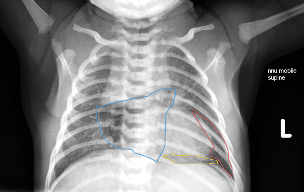
Concurrent Neonatal Pneumothorax, Pneumomediastinum, and Pneumopericardium: A Case Report of Multi-Compartment Air Leak Syndrome
When a routine neonatal check turns up a rare triple air leak without sending an infant into acute...

Empowering Women as Knowledge Creators through Open Education Initiatives
When Tri Astari addressed the Open Education Conference 2026 (OpenEd26) on October 27, 2026, she highlighted that increasing...

Changi Airport Upgrades iFerret AI to Detect 4 cm Runway Debris
Changi Airport is upgrading its iFerret runway surveillance network with advanced artificial intelligence to detect foreign objects as...