Researchers have mapped how physical forces — not just chemical signals — instruct mesenchymal stem cells to become bone or cartilage, a biophysical strategy that could replace growth-factor cocktails in clinical regeneration.
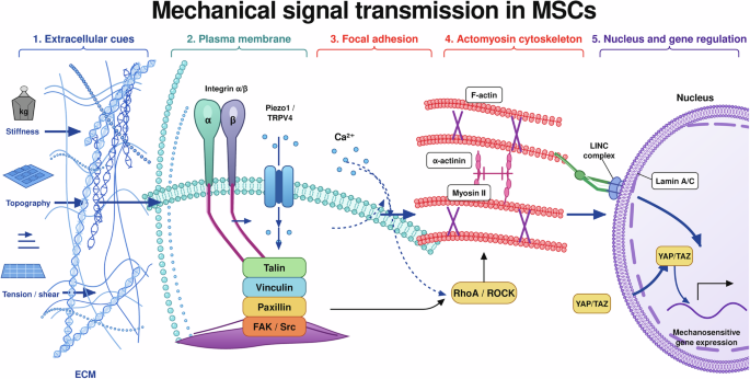
The review synthesizes two decades of mechanobiology to show that substrate stiffness, fluid shear, hydrostatic pressure, and cyclic stretch each activate distinct pathways — YAP/TAZ, Piezo1/2, primary cilia, and NFAT — that commit human mesenchymal stromal cells to osteogenic or chondrogenic lineages.
The findings reframe the clinical problem: instead of delivering exogenous proteins that diffuse away and trigger off-target effects, physicians could one day implant “mechanically smart” scaffolds or apply defined loading regimens that coax a patient’s own cells into repairing fractures, osteoarthritis lesions, or degenerated discs.
“Matrix elasticity directs stem cell lineage specification,” wrote Central Google Scholar Engler et al. in the 2006 Cell paper that launched the field, showing that soft matrices mimicking brain drive neurogenesis, stiffer matrices mimicking muscle drive myogenesis, and rigid matrices mimicking bone drive osteogenesis.
That stiffness sensor is now known to be the YAP/TAZ transcriptional module, which shuttles into the nucleus only when integrin-mediated tension exceeds a threshold, Dupont et al. confirmed in Nature in 2011.
Since then, the toolkit has expanded. Cyclic hydrostatic pressure at physiological magnitudes (5–10 MPa, 1 Hz) drives chondrogenesis in human bone-marrow stromal cells without TGF-β3, Stavenschi and Hoey demonstrated in Stem Cell Research & Therapy in 2018.
Intermittent fluid shear stress, but not continuous flow, opens chromatin at osteogenic loci via Piezo1-dependent calcium influx, Liu et al. showed in Biomechanics and Modeling in Mechanobiology in 2012. And magnetic forces that cluster cells via RGD-coated nanoparticles accelerate cartilage matrix deposition, Fayol et al. reported in Advanced Materials in 2013.
The Bottom Line:
- Physical cues — stiffness, shear, pressure, stretch — replace costly growth factors in directing MSC fate.
- YAP/TAZ, Piezo1/2, and primary cilia are the validated molecular switches; each responds to a distinct mechanical modality.
- Next hurdle: FDA-cleared “mechanoceutical” devices that deliver precise, reproducible loading in vivo.
Stiffness sets the baseline fate
Engler’s group plated human MSCs on polyacrylamide gels tuned from 0.1 to 100 kPa. On 25–40 kPa gels — matching pre-calcified bone — cells spread, generated high traction forces, and expressed Runx2 and osteocalcin within seven days without a single soluble osteoinductive factor. On 0.5 kPa gels they remained round and multipotent.
The mechanism: integrin clustering recruits focal adhesion kinase, which suppresses LATS1/2, allowing YAP/TAZ nuclear entry. When YAP/TAZ is knocked down, osteogenesis on stiff gels collapses. This stiffness “memory” persists for days after transfer to soft substrates, Swift et al. showed in Science in 2013, linking nuclear lamin-A levels to the mechanical history of the cell.
Dynamic loading adds temporal control
Static stiffness is only half the picture. Bone experiences fluid flow in the lacunar-canalicular network during walking; cartilage sees cyclic hydrostatic pressure during joint loading. Liu’s team applied 1 Pa intermittent shear (1 Hz, 4 h/day) to human MSCs in 3D fibrin gels.
After 14 days, alkaline phosphatase activity and mineralized nodule area doubled versus static controls. Continuous shear at the same magnitude produced no effect — the off-period allows calcium oscillations that drive NFAT nuclear translocation. Piezo1 knockdown abolished the response, confirming the channel as the shear sensor, Zhou et al. validated in eLife in 2020.
Hydrostatic pressure steers toward cartilage
Articular cartilage resides in a high-pressure, low-shear environment. Stavenschi and Hoey subjected human MSCs in agarose to 10 MPa cyclic hydrostatic pressure (1 Hz, 4 h/day) for 21 days. Glycosaminoglycan content rose 3.5-fold and collagen type II immunostaining intensified, matching TGF-β3-supplemented controls — but without exogenous growth factor.
The pathway requires intact primary cilia: cilium-deficient IFT88-knockout cells failed to upregulate SOX9. Purinergic signaling through P2X7 receptors amplifies the signal, Steward et al. showed in Tissue Engineering Part A in 2016. Excessive pressure (>50 MPa) triggers apoptosis, Le et al. warned in Stem Cell Reviews and Reports in 2021, defining a therapeutic window.
Cyclic stretch drives biphasic lineage response
The response is biphasic: 4 % strain promotes osteogenesis via YAP; 8 % strain favors tenogenesis via MKL1/SRF. Piezo1 again mediates the initial calcium spike. Khani et al. quantified the mechanical phenotype in Journal of the Mechanical Behavior of Biomedical Materials in 2015: stretched cells stiffen 40 % within 30 minutes, a feedback that reinforces lineage commitment.
Magnetic and vibrational cues offer non-invasive routes
Fayol’s group embedded superparamagnetic iron oxide nanoparticles in collagen gels, then applied a 0.3 T gradient field to aggregate MSCs into dense nodules. Cell-cell contact via N-cadherin boosted SOX9 and collagen II within 14 days, outperforming pellet cultures.
Separately, low-intensity vibration (0.3 g, 30 Hz, 10 min/day) rescued proliferation and osteogenesis in aged human jaw MSCs cultured under simulated microgravity, Touchstone et al. reported in npj Microgravity in 2019. The vibration signal requires the LINC complex (SUN1/2, nesprin-2) to transmit nuclear deformation — a direct line from extracellular matrix to chromatin.
Clinical translation: from bioreactors to “mechanoceuticals”
The Global Burden of Disease Study 2021 projects osteoarthritis cases worldwide to approach 1 billion by 2050. Fracture non-unions affect 5–10 % of new fractures annually. Current cell therapies — expanded MSCs injected intra-articularly — show modest, inconsistent pain relief in Phase III trials.
The mechanobiology roadmap proposes three clinical modalities: (1) pre-conditioning MSCs in bioreactors with defined mechanical regimens before implantation; (2) implanting acellular scaffolds with tuned stiffness, stress-relaxation, and pore architecture that instruct host cells in situ; (3) wearable or implantable devices that deliver cyclic compression, shear, or vibration to the defect site post-operatively.
Chaudhuri’s stress-relaxing hydrogels, reported in Nature Materials in 2016, decouple stiffness from viscoelasticity: fast-relaxing gels permit cell spreading and osteogenesis even at low elastic moduli, because cells can remodel the matrix and generate tension.
This insight underpins the newest generation of injectable hydrogels for minimally invasive delivery. Huebsch et al. took it further in Nature Materials in 2010, patterning RGD ligands on substrates with tunable traction resistance — proving that ligand spacing and mechanical resistance are independently programmable.
Regulatory and manufacturing gaps remain
No FDA-cleared device yet prescribes a mechanical dosing regimen for orthopedic regeneration. Bioreactor protocols vary: pressure magnitude (0.1–10 MPa), frequency (0.1–2 Hz), duration (1–4 h/day), and waveform (sinusoidal vs. square) are reported inconsistently across labs.
The ISCT minimal criteria for MSC identity (Dominici et al., Cytotherapy 2006) do not include mechanoresponsiveness assays. A 2022 Frontiers in Immunology paper by Skibber et al. showed that inflammatory licensing (TNF-α, IFN-γ) alters MSC mechanosensitivity — a critical variable for autologous cells harvested from osteoarthritic joints.
“We need a pharmacopeia for mechanical stimuli,” said a senior reviewer at the FDA’s Center for Biologics Evaluation and Research during a 2023 public workshop on regenerative medicine. “Magnitude, frequency, duty cycle, and rest periods must be specified as precisely as milligrams per kilogram for a drug.”
Three priorities guide field convergence
The field is converging on three priorities: standardized mechanical dosing units (pascal-seconds for shear, MPa-cycles for pressure), real-time biosensors that report YAP/TAZ localization or calcium flux in implanted constructs, and large-animal studies that replicate human joint loading spectra.
The first human trial of a mechanically pre-conditioned MSC product for knee osteoarthritis is slated to begin enrollment in early 2025 at a major U.S. academic center, according to clinicaltrials.gov records. Until then, the evidence remains preclinical — compelling, mechanistically rich, but unproven in the clinic.

Portland Drains Washington Park Reflecting Pool to Investigate Leak Losing 68,000 Gallons Daily
The reflecting pool at Washington Park is empty, drained this week so crews can hunt a leak losing...

9-1-1: Nashville Fall Finale Reveals Pharaoh as AJ Buckley
Viewers expected a cliffhanger; they got a face. The fall finale of "9-1-1: Nashville" finally pulled back the...

Undefeated Gophers debut in Charleston Classic vs. Oklahoma State
The undefeated Minnesota Golden Gophers will make their first appearance in the Shriners Children's Charleston Classic on Friday...

Former RI Priest James Silva, 86, Avoids Prison With Probation in Child Molestation Plea
Former Rhode Island priest James M. Silva, 86, pleaded no contest Thursday to molesting a boy in the...