
Guidelines say IVF pregnancies merit a closer look at the fetal heart. A new study suggests the standard look isn’t close enough.
Researchers at UPMC Harrisburg reviewed 355 low-risk IVF pregnancies and found a congenital heart defect rate of 10.3 percent — more than ten times the 0.8 percent baseline seen in the general population, Cureus reported. Three-quarters of the defects caught on a dedicated fetal echocardiogram had been missed on the detailed anatomy scan that current guidelines treat as sufficient.
“If a fetus is considered to have a greater than baseline risk of CHD, such as IVF, then a fetal echocardiogram is recommended in addition to the detailed fetal anatomical ultrasound,” the study authors wrote, citing American Heart Association and AIUM guidance. Their data argue that the “in addition” part is where the system breaks down.
“Nineteen of the 25 (76.0%) congenital heart defects discovered on fetal echocardiogram were not found on detailed obstetric ultrasound,” the UPMC team reported.
The retrospective cohort, covering deliveries from October 2016 through November 2021, excluded patients with other known risk factors — pregestational diabetes, chromosomal abnormalities, or a family history of heart defects — to isolate the IVF variable. Of the 30 total defects identified, 25 were spotted prenatally via echocardiogram: 19 left-to-right shunts, five left-heart obstructive lesions, and one cyanotic lesion. Another five surfaced only after birth.
A separate, larger study out of Europe paints a more complicated picture. Analyzing 2,877 fetuses referred for echocardiography over three years, researchers found no increase in major cardiac anomalies among ART pregnancies compared to natural conception, according to europepmc.org.
They did, however, report a significantly higher rate of mild cardiac anomalies — 51.4 percent versus 44.4 percent, p=0.03 — while noting that increased nuchal thickness and extra-cardiac anomalies were actually more common in the natural-conception group.
That contrast — tenfold increase versus no increase in major defects — lands squarely on how “defect” is defined and who gets scanned. The UPMC study screened a defined low-risk IVF cohort. The European study looked at a referred population already getting echocardiograms for “various reasons,” diluting the IVF signal with higher-risk indications.
Both studies agree on one operational reality: fetal echocardiograms demand specialized sonographers, pediatric cardiology oversight, and machine time that detailed anatomy scans already consume. “Many images are duplicated between the detailed fetal anatomical ultrasound and the fetal echocardiogram, with a significant strain on resources,” the UPMC authors said.
With IVF accounting for roughly 2 percent of U.S. live births as of 2018, the absolute number of affected families is small but not trivial. The UPMC data suggest that for every 100 low-risk IVF pregnancies, ten will carry a fetal heart defect — and in seven or eight of those cases, the routine anatomy scan will offer false reassurance.
Founded in 1873 as Harrisburg Hospital, UPMC Harrisburg performed the region’s first open-heart surgery in 1956 and its first percutaneous transluminal coronary angioplasty in 1980. The hospital joined the UPMC system in 2017 after operating for decades as the flagship of PinnacleHealth, and opened a dedicated Level III neonatal intensive care unit in 2019.
Prospective controlled studies are necessary to determine the true increase in disease incidence, the UPMC team concluded.
Worth a look
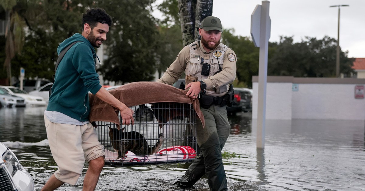
Central Florida Flood Crisis: 28 Inches of Rain, Record River Levels, Dozens Rescued
Central Florida’s Atlantic coast is waking up to a flood crisis that has already rewritten November rainfall records...

LA Council Tables Charter Amendments on Mayor’s Power After Timing Dispute
The Los Angeles City Council tabled two charter amendments Friday that would have weakened the mayor’s control over...

Peanut and Coterie Launch Village Fund With $200–$3,000 Grants for Mothers
Peanut and Coterie launched the Village Fund, committing a six-figure sum to grants of $200 to $3,000 for...
NYISO Warns of Statewide Power Shortfall as Early as 2033
"We've seen this happen in other states, other parts of the U.S., Louisiana, California, Texas. New York does...