A Low-Cost Training Tool for Rib Fracture Pain Management
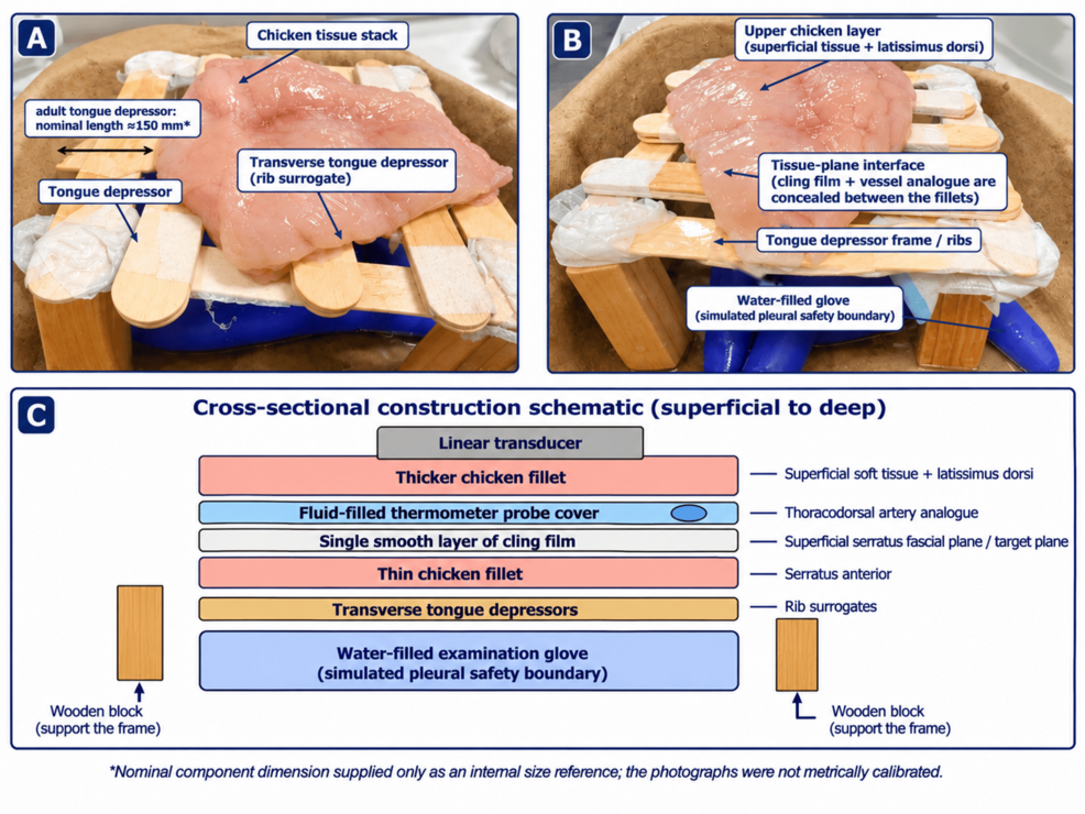
Researchers have developed a makeshift, low-cost simulator designed to help medical trainees practice a specific nerve block used to treat rib fracture pain, according to a report in Cureus. The device, a “part-task trainer,” uses everyday items like chicken fillets, wooden blocks, and tongue depressors to mimic the complex anatomy of the human chest wall. By allowing students to rehearse the precise needle movements required for a serratus anterior plane block (SAPB) without risking patient safety, the model aims to bridge the gap between classroom learning and real-world clinical practice.
The stakes for mastering this procedure are high. Rib fractures can cause debilitating pain that prevents patients from breathing deeply or coughing, which significantly increases the risk of respiratory complications. Effective pain management is critical; research published by PMC indicates that using ultrasound-guided SAPB can reduce postoperative pain, lower the need for supplemental opioids, and stabilize vital signs like heart rate and blood pressure in patients undergoing thoracoscopic surgery. However, performing the block requires a high level of skill, as clinicians must navigate near the pleura—the membrane surrounding the lungs—and avoid vital blood vessels.
The simulator addresses two primary safety concerns: accidental vascular puncture and pleural breach. The Cureus report details how the model includes a fluid-filled thermometer probe cover to act as a surrogate for the thoracodorsal artery. This allows trainees to practice identifying and avoiding the vessel during the injection. Additionally, a water-filled examination glove is positioned beneath the “ribs” to serve as a simulated pleural boundary. If a trainee’s needle trajectory is too deep, they receive immediate visual feedback by puncturing the glove, signaling an unsafe maneuver that, in a real patient, could lead to a pneumothorax (a collapsed lung).
Anatomy Replicated with Household Materials
The construction of the phantom is a study in pragmatic medical education. Rather than using expensive, high-fidelity synthetic models, the developers opted for biological and household substitutes that provide realistic tactile feedback. The “tissue” is composed of two layers of chicken breast, with a single layer of cling film placed between them to represent the fascial plane where the local anesthetic is actually deposited. This plane is the target for “hydrodissection,” a technique where fluid is injected to separate tissue layers and confirm the correct placement.
To provide a rigid structure, the model uses four wooden stacking blocks as legs and standard wooden tongue depressors to serve as the ribs. The depth of these “ribs” is calibrated to approximately 18–25 mm, while the target superficial plane sits roughly 8–12 mm below the skin. While these dimensions are approximations used for a proof-of-concept build, they offer a functional environment for practicing “in-plane” needle visualization, where the clinician must track the entire length of the needle on an ultrasound screen.
The Path from Simulation to Patient Care
This device is not intended to be a standalone certification tool. Instead, the developers propose a structured seven-step pathway that uses the simulator as a gatekeeper. In this model, a trainee must demonstrate proficiency on the phantom before they are permitted to move toward supervised practice on actual patients. This staged approach is designed to build the necessary hand-eye coordination and sonographic recognition required for regional anesthesia.
The transition from simulation to the bedside is a critical moment in medical training. The Cureus study notes that while the model is a starting point for preclinical preparation, it does not replace the need for formal clinical assessment or the expertise of a supervisor. The goal is to ensure that by the time a trainee reaches a patient, the fundamental mechanics of the block—identifying the serratus anterior muscle and avoiding the pleural boundary—have already become second nature.
While the clinical benefits of the SAPB are well-documented, the medical community continues to refine how these skills are taught. The development of low-cost trainers like this one suggests a shift toward more accessible, repeatable training modules that can be deployed in various clinical settings, from academic hospitals to emergency departments, ensuring that the next generation of clinicians can manage acute pain with greater precision and lower risk.
Keep reading

Punjab Police Deport Wanted Gangster Gurwinder alias Afridi From Ethiopia After 10-Month Manhunt
Gurwinder, widely known as Afridi, landed at Delhi’s Indira Gandhi International Airport early Sunday morning, marking the end...
High-Dose Flu Vaccine Linked to Lower Alzheimer’s Risk in Older Adults
High-dose influenza vaccines may reduce Alzheimer's risk by nearly 55% A higher dose of the annual flu shot...

Locavore NXT in Bali tests zero-import dining and sustainability
The radical pursuit of zero imports in Bali In the heart of Ubud, a culinary experiment is testing...

PAGASA: Three Weather Systems to Bring Rains and Cloudy Skies Across PH on Sunday
Three weather systems bring rain and flood risks to the Philippines Residents across much of the Philippines should...