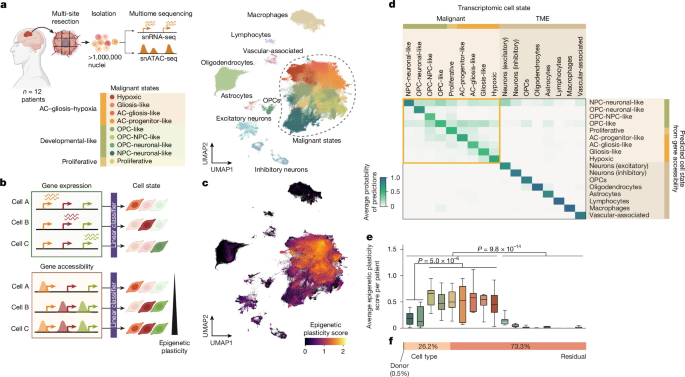
For years, glioblastoma has been described as a chaotic disease. The most common and lethal brain cancer in adults contains a shifting mixture of cancer cell states that seemed to obey no consistent pattern — one reason treatment so often fails and the cancer returns. Now, a large collaborative study published in Nature argues that this apparent randomness is an illusion, and that the tumor’s cells follow predictable structural rules as they grow.
Researchers at the Wellcome Sanger Institute, the European Molecular Biology Laboratory, the German Cancer Research Center, and the Cancer Research UK Cambridge Institute mapped more than one million individual cells across glioblastoma tumors from 12 patients, taking samples from different regions of each tumor at Cambridge University Hospitals NHS Foundation Trust. What they found challenges a long-held assumption that glioblastoma cell states are too dynamic and disordered to be systematically targeted: the same sequence of events repeats from one patient to the next.
One million nuclei, 57 tumor sites, and a hidden order
The scale of the dataset is what makes the finding hard to dismiss. The team processed sites across 12 tumors, isolating 1,025,329 nuclei and profiling them with paired single-nucleus RNA and chromatin accessibility sequencing, alongside spatial transcriptomic maps of adjacent tissue sections. Only samples with an RNA integrity number above 7 made the cut, and each tumor was sampled at multiple sites — superior, anterior, posterior, and middle regions where possible — so the researchers could see how cell states arrange themselves in space, not just in a test tube.

To make sense of this complexity, the team built a deep-learning method called scDORI, which unifies dimensionality reduction and gene-regulatory network inference so that each latent factor carries regulatory meaning. Trained on 95% of the atlas and evaluated on the held-out 5%, it learned 50 latent “Topics” — signed, enhancer-mediated networks linking co-accessible chromatin peaks, co-expressed genes, and their predicted activating and repressing regulators. Thirty-one of those Topics were active enough to analyze.
The payoff came when the team integrated the single-cell data with spatial transcriptomic data. “By integrating the single-cell data with spatial transcriptomic data, we found that there is a meticulous and very specific spatial architecture of cancer cell states,” said Bayraktar of the study team. “And this was present in every single glioblastoma tumor in every single patient we studied.” The pattern held when the researchers grew patient-derived tumor cells into three-dimensional brain organoids in the lab, confirming the trajectory matched what happens inside real tumors.
Why plasticity makes glioblastoma so hard to kill
The stakes are stark. Around 3,200 people are diagnosed with glioblastoma every year in the United Kingdom. Average survival is 12 to 18 months, and only 4% of those diagnosed survive five years or more. Standard treatment — surgery followed by chemotherapy and radiotherapy — is difficult because the tumors sit in the brain and invade multiple regions, which can make surgery impossible.
Even when treatment destroys most tumor cells, a highly dynamic population of cancer cell states lets some cells survive, leading to drug resistance and the cancer’s re-emergence. The study quantified this plasticity directly: researchers measured the degree of decoupling between chromatin accessibility and transcription at marker genes, finding that accessible chromatin at loci not yet transcribed indicates a “primed” configuration poised to switch into a different identity. Cells whose accessibility was compatible with several transcriptional states scored as more plastic — exactly the cells that let the tumor adapt around therapy.
The new atlas shows that adaptability is not random. “A tumor generates an almost endless number of variations on its own regulatory program,” said Stegle of EMBL Heidelberg and DKFZ. “Instead of seeing this heterogeneity only as an obstacle, we can use AI to learn from it and extract the rules that determine how cells choose their fate.”
From mapping to reprogramming: the MYT1L test
The predictable trajectory has a practical implication: if cell states follow rules, those rules can potentially be bent. The researchers identified “safeguard” regulators — transcription factors whose repressive links reach across multiple alternative cell states — and tested one of them, MYT1L, in patient-derived brain tumor cells.
“We could perhaps reprogram them, and maybe push them into a state that is less malignant, less dangerous,” as the team put it. Overexpressing MYT1L reduced cellular plasticity and tumor malignancy in those experiments. The researchers are now applying the same framework to other cancers and brain disorders; clinical applications may still be some way off, but the atlas and the AI tool have already surfaced new regulatory sequences that could drive specific cell states in a tumor-specific setting.
What the study does not claim is a cure. The organoid and cell-line experiments are early, and the authors themselves frame the work as a framework for spotting common patterns at the deep molecular level. But the shift in framing matters: a disease long treated as unpredictable now has a measurable architecture, and that is the kind of finding that eventually becomes a target.
Worth a look
Google Runs AI Model in Orbit, Transmits Poem to Earth
Google confirmed on October 30 that its Project Suncatcher prototype has run an artificial intelligence model in orbit...

US-funded newborn vaccine trial in Guinea-Bissau cancelled after WHO objections
A US-funded vaccine trial on newborns in Guinea-Bissau has been cancelled after the World Health Organization raised alarms...

Galway City Ring Road: Cycling Campaigner Withdraws Challenge, Paving Way for Public Consultation
The last legal obstacle blocking the Galway City Ring Road fell away this week when cycling campaigner Shane...

PAGASA Forecasts Scattered Rains and Thunderstorms Over Parts of PH
The Philippine Atmospheric, Geophysical and Astronomical Services Administration (PAGASA) has forecast scattered rains and thunderstorms across parts of...