
Engineers at the Massachusetts Institute of Technology have invented a high-speed microscope capable of capturing electrical activity across the entire brain of an intact organism according to MIT. The technology allows researchers to track neuronal voltage signals across the whole brain of larval zebrafish at a rate of once every five milliseconds, an advance reported in Nature Methods.
MIT Engineers Develop High-Speed Microscope for Whole-Brain Voltage Imaging
Neurons communicate by generating electrical impulses that travel through vast interconnected networks controlling behavior, sensory perception, memory formation, and movement. Capturing these rapid signals simultaneously across large brain regions has historically presented significant technical hurdles. The newly developed system enables scientists to resolve individual voltage spikes across distributed neural circuits in real time.
Overcoming the Limitations of Calcium Imaging
Traditional studies of neuronal activity frequently rely on calcium imaging, a method that measures changes in intracellular calcium levels flowing into cells after an electrical impulse fires. While useful as a proxy for neural activity, calcium imaging operates on a much slower timescale.
Former MIT research scientist Jie Zhang noted that calcium imaging is inherently slow, capturing activity on the order of seconds or minutes. Such speeds fail to capture high-speed neural spikes. To bypass this limitation, the MIT team sought a method capable of millisecond-scale resolution across a large volumetric space such as the entire brain.
Modifying Light-Sheet Microscopy With Remote Refocusing
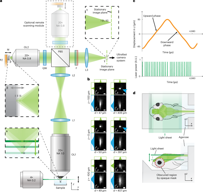
To achieve the necessary resolution and speed, the research team modified a standard light-sheet microscope, which uses a sheet of laser light to illuminate a thin sample slice and generate three-dimensional images of large volumes by scanning multiple layers in sequence.
To accelerate the imaging process to the millisecond scale, researchers increased the image acquisition speed of the camera and boosted the scanning speed using a technique known as remote refocusing. This setup enabled the system to scan the entire zebrafish brain 200 times per second, or once every five milliseconds, supporting volumetric imaging rates above 200 volumes per second across a 200-micrometer axial range.
To test the microscope, researchers engineered neurons in larval zebrafish to express a voltage indicator called Positron2-Kv. The indicator produced signals in neurons distributed throughout the brain, with approximately one quarter of the neurons exhibiting acceptable signals.
Implications for Neurotechnology and Systems Neuroscience
The research aims to help neuroscientists investigate how widely separated groups of neurons coordinate to produce behaviors, sensory responses, and other complex functions.
All of the parts of the brain are connected together, so if you want to truly understand the brain, you have to understand how all the neurons work together as an emergent whole,
said senior author Wiley, the Y. Eva Tan Professor in Neurotechnology at MIT. Boyden is also a professor of biological engineering, media arts and sciences, and brain and cognitive sciences, alongside holding affiliations with MIT’s McGovern Institute for Brain Research, Yang Tan Collective, and the Koch Institute for Integrative Cancer Research.
The study was authored by senior author Ed Boyden alongside lead authors Zeguan Wang, a former J. Douglas Tan Postdoctoral Fellow who earned his PhD in 2024, and former MIT research scientist Jie Zhang.
Worth a look

Moon to Occult Jupiter Over Eastern North America on October 6
Skywatchers across much of eastern North America can watch a rare lunar occultation early Tuesday morning as a...

Saturn Reaches Closest Approach to Earth for the Year
Saturn stands roughly 784 million miles from Earth as opposition positions the ringed giant at its closest annual...

Researchers Identify Candidate Second-Generation Planet Orbiting White Dwarf
Circling its parent dead star every 4.4 days, a Jupiter-sized gas giant orbits the white dwarf HS 0209+0832...

UK Proposes Norfolk Housing Levy to Unlock Stalled Homes
Environment Secretary Angela Eagle launched a public consultation on 17 September for a Norfolk pilot scheme requiring housing...