
Coronary inflammation measured via non-invasive imaging significantly improves long-term cardiovascular risk assessment following percutaneous coronary intervention. A retrospective study of 490 patients evaluated pericoronary adipose tissue attenuation to refine risk stratification beyond traditional plaque characteristics and calcium scores.
Traditional cardiovascular risk assessments often miss vulnerable patients before they experience a cardiac event. This gap in early detection has prompted researchers to investigate inflammatory markers visualised through standard imaging protocols to better capture vascular disease activity.
Pericoronary Adipose Tissue Attenuation and Post-PCI Risk
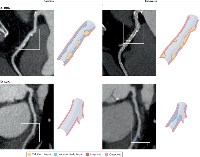
A retrospective study evaluating pericoronary adipose tissue (PCAT) attenuation as visualised by coronary computed tomography angiography (cCTA) examined its prognostic value in patients undergoing percutaneous coronary intervention with current-generation drug-eluting stents. Researchers analysed data from 490 patients who received cCTA prior to their procedures, assessing adverse plaque characteristics, calcified plaque burden, and PCAT attenuation specifically in the proximal right coronary artery.
The study tracked a patient-oriented composite endpoint defined as a combination of cardiovascular death, non-fatal myocardial infarction, revascularisation, and stroke. Over a median follow-up period of 1,540 days, 77 patients experienced this composite endpoint. These individuals exhibited significantly higher proximal right coronary artery PCAT attenuation values of -76.3±6.4 HU, compared with -82.5±8.1 HU for patients without events.
Multivariable Analysis and Risk Reclassification Thresholds
Multivariable analysis confirmed that several imaging markers were independently associated with adverse outcomes following intervention. Specifically, the presence of adverse plaque carried a hazard ratio of 2.05, greater calcified plaque burden carried a hazard ratio of 1.04, and higher proximal right coronary artery PCAT attenuation carried a hazard ratio of 2.20. Patients with attenuation values at or above -79.9 HU demonstrated a 3.9-fold higher incidence of adverse outcomes compared to patients below that threshold.
When researchers incorporated these PCAT attenuation measurements into risk models alongside traditional cardiovascular risk factors and cCTA-based plaque assessments, predictive accuracy and patient risk reclassification improved significantly.
Intersecting Metabolic Disease and Coronary Calcification
Type 2 diabetes mellitus and stable ischemic heart disease frequently coexist, defining a population highly vulnerable to adverse cardiovascular events. Chronic hyperglycemia contributes to endothelial dysfunction, oxidative stress, and the formation of advanced glycation end products, which promote vascular inflammation and osteogenic differentiation of vascular smooth muscle cells.
Between January 2016 and December 2022, a retrospective screening evaluated 4,733 patients with type 2 diabetes mellitus and stable ischemic heart disease who underwent coronary computed tomography angiography at a single institution. After applying exclusion criteria—such as a coronary artery calcium score of zero, acute coronary syndrome, chronic heart failure, left main disease, or severe renal dysfunction—the final analysis included 3,991 patients.
Clinical Integration and Future Platform Development
As diagnostic imaging tools evolve, artificial intelligence applications are expanding the utility of routine cardiovascular scans. According to recent studies, AI-enabled analysis of coronary CT angiography demonstrates close agreement with expert readers and intravascular ultrasonography, with potential prognostic value for predicting future myocardial infarction (Lin et al., 2022; Föllmer et al., 2024).
These developments highlight a broader shift in cardiovascular medicine supported by international consensus statements and large-scale clinical trials like SCOT-HEART and DISCHARGE, which have progressively shaped diagnostic imaging guidelines and reimbursement frameworks across Europe and the United States.
Keep reading

Brain Health Improvement Relies on Mental Stimulation and Hearing Care
Improving long-term brain health and cognitive vitality involves a combination of science-backed lifestyle habits, targeted daily practices, and...
Ketogenic Diet Reduces Liver Fat Better Than Other Diets
A randomized clinical trial published in Cell Metabolism reveals that adults with obesity and prediabetes who lost about...

Nepal Authorities Initiate Mass Cremations as Flood Death Toll Hits 800
Following a catastrophic glacial collapse along the Nepal-China border on August 26, 2026, the death toll from widespread...

Breast Cancer Survivors Face Long-Term Physical and Emotional Challenges
Breast cancer survivors often face profound physical, emotional, and practical challenges after treatment ends.