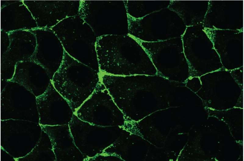
Researchers at the Whitehead Institute have recreated planar cell polarity in engineered epithelial cells, discovering that collective cell migration acts as a physical cue to establish coordinated orientation across cell sheets. Published on October 15 in the Proceedings of the National Academy of Sciences, the findings identify the junctional protein CELSR as a key driver of this symmetry-breaking process.
From the microscopic sensory structures of the inner ear to the protective hairs on our skin, building functional tissues requires individual cells to know which direction they are facing. Cells must coordinate their orientation across sheets that cover the body’s surfaces and line its internal organs. When this spatial coordination breaks down, tissues often fail to form or function properly. Disruptions to planar cell polarity are linked to serious developmental disorders, including spina bifida, craniorachischisis, hearing loss, and chronic lung disease.
Scientists have long understood many of the proteins involved in planar cell polarity, but a fundamental mystery remained: what initially tells unorganized cells which direction to face? While signaling gradients like WNT proteins and mechanical forces help maintain polarity in already organized tissues, catching the exact moment of initiation inside a developing animal has proved exceedingly difficult.
Recreating Tissue Polarity in Whitehead Institute Cell Cultures
To solve that observational puzzle, a research team working in the laboratory of Whitehead Institute Member Pulin Li rebuilt the developmental process from the bottom up. Using petri dishes, the researchers cultured engineered epithelial cells that initially lacked any planar polarity. They fluorescently labeled the proteins responsible for establishing polarity so they could track cellular movements in real time.
During the experiment, the team observed an unexpected mechanism. When the cells were induced to migrate collectively, junctional proteins containing CELSR became heavily enriched along the front-to-back axis of the movement. That molecular asymmetry successfully aligned across neighboring cells and propagated continuously backward through the sheet, transforming an unorganized collection of cells into a coordinated tissue.
We’ve identified a cue, and we’ve also found what we think is the protein responsible for actually reading that signal.
Wallach, via Newswise
That observation sheds light on a classic symmetry-breaking event in developmental biology, demonstrating how collective cell migration can provide the initial spatial cue that sparks a molecular pattern.
Dissecting the Role of CELSR and Individual Cell Responsiveness
The controlled in vitro system allowed the researchers to test competing hypotheses about how polarity spreads across a tissue. One possibility was that each cell independently interprets the directional cue; another was that a small subset of cells senses the signal first and relays instructions to their neighbors. The findings supported the first model, showing that individual cells directly interpret migration cues and stay responsive to them.
The experiments also clarified the function of CELSR, a protein that forms connections at junctions between adjacent cells. Core planar cell polarity typically involves CELSR alongside two other protein families: Frizzled and Van Gogh-like. Surprisingly, the emergence of CELSR polarity occurred without requiring reciprocal interactions with Frizzled or Van Gogh-like proteins, pointing to CELSR as an independent driver of polarity initiation.
Being able to recreate this process in cell culture lets us separate pieces that are extremely difficult to separate in an animal and watch the pattern form in real time.
Leah Wallach, graduate student in the Li lab, via Newswise
Implications for Engineered Tissues and Future Developmental Biology
Traditionally, developmental biology views cell movement as a downstream outcome of molecular patterning programs, where cells receive spatial instructions first and move accordingly. This study proves that the relationship can operate in reverse, with collective behavior generating spatial information and molecular order.
Coordinating the orientation of cells is essential for the function of tissues such as the airway and inner ear, yet remains difficult to achieve in engineered tissues.
Pulin Li, Whitehead Institute Member, via Newswise
Epithelial lumen formation, for instance, is fundamental to organ development, yet how apical surfaces first emerge remains unclear according to separate findings reported by Ghosh, Martin, and Chen. For now, the Whitehead Institute team has established a reliable laboratory model to trigger cell directionality and observe the emergence of organized tissues from the molecular level upward.
Keep reading

Thwaites Glacier Melts Faster Than Expected. Sea Levels Could Double by 2100
Thwaites Glacier in West Antarctica is melting faster than expected, with a new model showing sea level rise...
Japanese Researchers Guide Vortices via Atomic Steps
Under intermediate magnetic fields, vortices traveled more than 1,000 times more easily along surface steps just one atom...

NASA Completes Optical Alignments on Nancy Grace Roman Space Telescope
Following an August 30 launch, Nancy Grace Roman Space Telescope team completed rigorous in-space optical alignments, achieving a...

University of Queensland Researchers Propose Omic Technologies to Develop Heat-Adapted Chickpeas
Researchers at the University of Queensland have proposed a staged approach using new omic technologies to accelerate the...