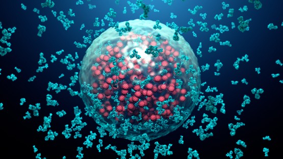
Researchers have developed new continuous-time simulation frameworks and optical imaging methods to track real-time cell metabolism and verify super-resolution fluorescence microscopy protocols. These advances improve how scientists study complex three-dimensional tissue structures and cancer metabolism.
Scientists Design Faster Imaging to Observe Cell Metabolism
Scientists designed a faster form of two-photon fluorescence imaging to observe cell metabolism in real time, a technique that could speed up screening for new therapeutic treatments. The findings were published Sept. 4 in Science Advances. Former postdoctoral researcher Lu Ling, Ph.D. ’20 and doctoral student Jack Crowley, M.S. ’22 share the role of co-lead author.
While conducting research in the laboratory of Claudia Fischbach—who serves as the James M. and Marsha McCormick Family Director of the Meinig School of Biomedical Engineering and the Stanley Bryer 1946 Professor of Biomedical Engineering within the Cornell Duffield College of Engineering—Ling launched the initiative. Researchers wanted a better way to monitor cancer metabolism, a process that changes within minutes or hours. Traditional methods lacked the tools required to track individual cell dynamics over extended periods.
“The idea was, can we do something better than the standard methods?”
Ling, co-lead author via Cornell
“We want to look at cancer metabolism, but it’s a very dynamic process. Everything can happen within minutes or hours, so there’s not really a good way to track each cell’s dynamics with the tools we had at that time,”
Ling, co-lead author via Cornell
Researchers Compare FPRM with Fluorescence Lifetime Imaging
Warren Zipfel, an associate professor of biomedical engineering in Duffield Engineering who focuses on applying optical microscopy to biomedical investigations, was contacted by Fischbach and Ling. Fluorescence lifetime imaging microscopy (FLIM), a conventional approach for tracking metabolism via cellular NADH fluorescence, particularly captured Fischbach’s interest. NADH is a small molecule that transports raw energy harvested from food directly into cellular machinery that produces power. FLIM works by exciting molecules and measuring how long they stay excited before releasing energy as fluorescent light, where the time delay of detected photons reveals details about local environment and behavior.
Instead, the team devised two-photon fluorescence polarization ratiometric microscopy (FPRM), an approach wherein a laser-scanning microscope directs polarized, focused light across cells while employing a polarizing beam splitter to simultaneously route the resulting fluorescence into two detectors. This provides a measure of how fast the emitting molecules are rotating.
Simulation Frameworks and Python Libraries Power Fluorescence Microscopy
Fluorescence lifetime imaging measures temporal decay after excitation to quantify a fluorophore’s local environment and molecular interactions, reporting changes associated with pH, ion concentration, viscosity, oxygenation, cellular metabolism, and FRET. Experimental measurements from supported acquisition systems are imported and preprocessed through detector-aware data-handling pathways, while simulation frameworks generate controlled fluorescence decays with known parameters and configurable acquisition and noise characteristics.
In the course of a separate computational imaging investigation, the PyFLI simulation framework served as underlying research infrastructure, enabling Erbas and colleagues to produce a collection of 1.6 million simulated fluorescence-lifetime signals to study low-precision recurrent neural-network inference. Continuous integration runs test suites on Ubuntu, Windows, and macOS with Python 3.11.
In another effort, researchers introduced MUFASA (Multi-Protocol Unified Fluorescence-based Advanced Simulation Algorithm), which functions as a physically grounded, continuous-time simulator for multiple super-resolution fluorescence microscopy techniques. By modeling fluorophore dynamics using continuous-time Markov chains, MUFASA yields realistic photon emission behavior across Single Molecule Localization Microscopy and fluorescence fluctuation-based settings, supporting individual emitters, structure-level simulations, photophysical transitions, photobleaching, and camera properties.
Applications Expand into Three-Dimensional Tissue Systems
“So, any opportunity to provide new measurement strategies, especially ones that are simpler and lower cost, means that more people can envision diverse experiments that watch dynamic responses in intricate three-dimensional tissue-engineered structures. And I think that is going to be amazing for research more broadly.”
Jack Crowley, co-lead author via Cornell
MUFASA also introduces a novel validation metric based on the 1-Wasserstein distance between simulated and experimental photon-count distributions, estimates key photophysical parameters like molar extinction coefficients, and suggests optimal light-source power ranges from fluctuation data using an intuitive Python-based graphical interface designed for biologists, physicists, microscopists, and computational imaging engineers.
Keep reading

Researchers Map Immune Cells to Reveal Biological Aging Trajectories
Researchers analyzing millions of immune cells from global cohorts have discovered that the ratio between two white blood...

Louisa Nicola Outlines Lifestyle Changes to Prevent Alzheimer’s
Neuroscientist Louisa Nicola argues that up to 95 percent of Alzheimer’s cases are preventable through lifestyle choices, pointing...

Marking World Mental Health Day 2026, the Irish government announced an €11 million funding package
Marking World Mental Health Day 2026, the Irish government announced an €11 million funding package to expand youth...

CU Anschutz Study: Horseback Riding Cuts Hyperactivity in Autistic Youth
Therapeutic horseback riding cuts hyperactivity in children with autism and co-occurring psychiatric conditions, according to new research published...